Explore
 Validate
 Learn
Learn Western blot
Antibody data
- Antibody Data
- Antigen structure
- References [9]
- Comments [0]
- Validations
- Western blot [6]
- Immunocytochemistry [1]
- Immunoprecipitation [1]
- Immunohistochemistry [2]
Submit
Validation data
Reference
Comment
Report error
- Product number
- GTX114008 - Provider product page

- Provider
- GeneTex
- Proper citation
- GeneTex Cat#GTX114008, RRID:AB_2036353
- Product name
- Bmi1 antibody
- Antibody type
- Polyclonal
- Reactivity
- Human, Mouse
- Host
- Rabbit
Submitted references CSC-3436 inhibits TWIST-induced epithelial-mesenchymal transition via the suppression of Twist/Bmi1/Akt pathway in head and neck squamous cell carcinoma.
MDM2 Degrades Deacetylated Nucleolin Through Ubiquitination to Promote Glioma Stem-Like Cell Enrichment for Chemotherapeutic Resistance.
NLRP3 inflammasome activation promotes inflammation-induced carcinogenesis in head and neck squamous cell carcinoma.
Endothelial Cells Promote Formation of Medulloblastoma Stem-Like Cells via Notch Pathway Activation.
Stress stimuli induce cancer-stemness gene expression via Sp1 activation leading to therapeutic resistance in glioblastoma.
Depletion of p21-activated kinase 1 up-regulates the immune system of APC∆14/+ mice and inhibits intestinal tumorigenesis.
Transcription factor SPZ1 promotes TWIST-mediated epithelial-mesenchymal transition and oncogenesis in human liver cancer.
Suberoylanilide hydroxamic acid represses glioma stem-like cells.
Identification of thiostrepton as a novel therapeutic agent that targets human colon cancer stem cells.
Lai YJ, Yu WN, Kuo SC, Ho CT, Hung CM, Way TD, Chen CT
Journal of cellular physiology 2019 Jun;234(6):9118-9129
Journal of cellular physiology 2019 Jun;234(6):9118-9129
MDM2 Degrades Deacetylated Nucleolin Through Ubiquitination to Promote Glioma Stem-Like Cell Enrichment for Chemotherapeutic Resistance.
Ko CY, Lin CH, Chuang JY, Chang WC, Hsu TI
Molecular neurobiology 2018 Apr;55(4):3211-3223
Molecular neurobiology 2018 Apr;55(4):3211-3223
NLRP3 inflammasome activation promotes inflammation-induced carcinogenesis in head and neck squamous cell carcinoma.
Huang CF, Chen L, Li YC, Wu L, Yu GT, Zhang WF, Sun ZJ
Journal of experimental & clinical cancer research : CR 2017 Sep 2;36(1):116
Journal of experimental & clinical cancer research : CR 2017 Sep 2;36(1):116
Endothelial Cells Promote Formation of Medulloblastoma Stem-Like Cells via Notch Pathway Activation.
Wang Y, Wang Y, Chen H, Liang Q
Journal of molecular neuroscience : MN 2017 Oct;63(2):152-158
Journal of molecular neuroscience : MN 2017 Oct;63(2):152-158
Stress stimuli induce cancer-stemness gene expression via Sp1 activation leading to therapeutic resistance in glioblastoma.
Chang KY, Huang CT, Hsu TI, Hsu CC, Liu JJ, Chuang CK, Hung JJ, Chang WC, Tsai KK, Chuang JY
Biochemical and biophysical research communications 2017 Nov 4;493(1):14-19
Biochemical and biophysical research communications 2017 Nov 4;493(1):14-19
Depletion of p21-activated kinase 1 up-regulates the immune system of APC∆14/+ mice and inhibits intestinal tumorigenesis.
Huynh N, Wang K, Yim M, Dumesny CJ, Sandrin MS, Baldwin GS, Nikfarjam M, He H
BMC cancer 2017 Jun 19;17(1):431
BMC cancer 2017 Jun 19;17(1):431
Transcription factor SPZ1 promotes TWIST-mediated epithelial-mesenchymal transition and oncogenesis in human liver cancer.
Wang LT, Chiou SS, Chai CY, Hsi E, Chiang CM, Huang SK, Wang SN, Yokoyama KK, Hsu SH
Oncogene 2017 Aug;36(31):4405-4414
Oncogene 2017 Aug;36(31):4405-4414
Suberoylanilide hydroxamic acid represses glioma stem-like cells.
Hsu CC, Chang WC, Hsu TI, Liu JJ, Yeh SH, Wang JY, Liou JP, Ko CY, Chang KY, Chuang JY
Journal of biomedical science 2016 Nov 18;23(1):81
Journal of biomedical science 2016 Nov 18;23(1):81
Identification of thiostrepton as a novel therapeutic agent that targets human colon cancer stem cells.
Ju SY, Huang CY, Huang WC, Su Y
Cell death & disease 2015 Jul 2;6(7):e1801
Cell death & disease 2015 Jul 2;6(7):e1801
No comments: Submit comment
Supportive validation
- Submitted by
- GeneTex (provider)
- Main image
- Experimental details
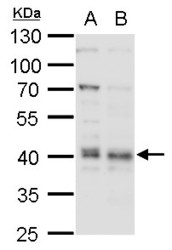
- Sample (30 ?g of whole cell lysate) A: H1299 10% SDS PAGE GTX114008 diluted at 1:1000 The HRP-conjugated anti-rabbit IgG antibody (GTX213110-01) was used to detect the primary antibody.

- Submitted by
- GeneTex (provider)
- Main image
- Experimental details
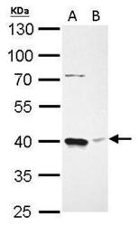
- Bmi1 antibody detects BMI1 protein by western blot analysis.A. 30 ?g THP-1 whole cell lysate/extractB. 30 ?g HL-60 whole cell lysate/extract10% SDS-PAGEBmi1 antibody (GTX114008) dilution: 1:1000 The HRP-conjugated anti-rabbit IgG antibody (GTX213110-01) was used to detect the primary antibody.

- Submitted by
- GeneTex (provider)
- Main image
- Experimental details
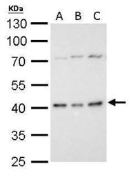
- Bmi1 antibody detects BMI1 protein by western blot analysis.A. 30 ?g C8D30 whole cell lysate/extractB. 30 ?g BCL-1 whole cell lysate/extractC. 30 ?g Raw264.7 whole cell lysate/extract10% SDS-PAGEBmi1 antibody (GTX114008) dilution: 1:1000 The HRP-conjugated anti-rabbit IgG antibody (GTX213110-01) was used to detect the primary antibody.

- Submitted by
- GeneTex (provider)
- Main image
- Experimental details
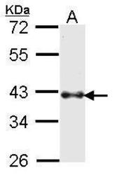
- Bmi1 antibody detects Bmi1 protein by western blot analysis. A. 30 ?g K562 whole cell lysate/extract B. 30 ?g THP-1 whole cell lysate/extract10% SDS-PAGEBmi1 antibody (GTX114008) dilution: 1:1000 The HRP-conjugated anti-rabbit IgG antibody (GTX213110-01) was used to detect the primary antibody.

- Submitted by
- GeneTex (provider)
- Main image
- Experimental details
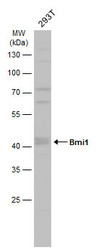
- Whole cell extract (30 ?g) was separated by 10% SDS-PAGE, and the membrane was blotted with Bmi1 antibody (GTX114008) diluted at 1:1000. The HRP-conjugated anti-rabbit IgG antibody (GTX213110-01) was used to detect the primary antibody.
- Submitted by
- GeneTex (provider)
- Main image
- Experimental details
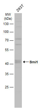
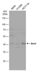
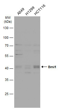
- Various whole cell extracts (30 ?g) were separated by 10% SDS-PAGE, and the membrane was blotted with Bmi1 antibody (GTX114008) diluted at 1:1000. The HRP-conjugated anti-rabbit IgG antibody (GTX213110-01) was used to detect the primary antibody.
Supportive validation
- Submitted by
- GeneTex (provider)
- Main image
- Experimental details
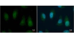
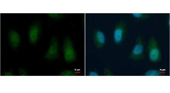
- BMI1 antibody [N1C3-2] detects BMI1 protein by immunofluorescent analysis. Sample: HeLa cells were fixed in 4% paraformaldehyde for 15 min. Green: BMI1 protein stained by BMI1 antibody (GTX114008) diluted at 1:1000. Blue: Hoechst 33342 staining. Scale bar = 10 £gm.
Supportive validation
- Submitted by
- GeneTex (provider)
- Main image

- Experimental details
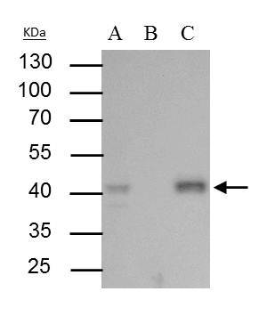
- Bmi1 antibody immunoprecipitates Bmi1 protein in IP experiments. IP Sample: 293T whole cell lysate/extract A : 30 £gg whole cell lysate/extract of Bmi1 protein expressing 293T cells B : Control with 2.5 £gg of pre-immune rabbit IgG C : Immunoprecipitation of Bmi1 protein by 2.5 £gg of Bmi1 antibody (GTX114008) 10% SDS-PAGE The immunoprecipitated Bmi1 protein was detected by Bmi1 antibody (GTX114008) diluted at 1 : 1000. EasyBlot anti-rabbit IgG (HRP) (GTX221666-01) was used as a secondary reagent.
Supportive validation
- Submitted by
- GeneTex (provider)
- Main image
- Experimental details
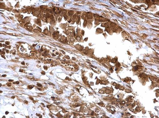
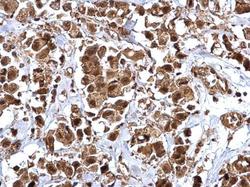
- Bmi1 antibody detects Bmi1 protein at cytosol and nucleus on human breast carcinoma by immunohistochemical analysis. Sample: Paraffin-embedded human breast carcinoma. Bmi1 antibody (GTX114008) dilution: 1:500.
- Submitted by
- GeneTex (provider)
- Main image

- Experimental details
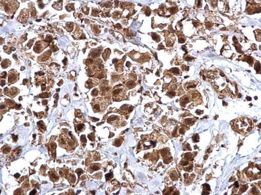
- Bmi1 antibody detects Bmi1 protein at cytosol and nucleus on human ovarian carcinoma by immunohistochemical analysis. Sample: Paraffin-embedded human ovarian carcinoma. Bmi1 antibody (GTX114008) dilution: 1:500.