 Explore
 Validate
 Learn
Learn Western blot
 ELISA
ELISAAntibody data
- Antibody Data
- Antigen structure
- References [0]
- Comments [0]
- Validations
- Western blot [2]
- Immunocytochemistry [1]
Submit
Validation data
Reference
Comment
Report error
- Product number
- GTX45791 - Provider product page

- Provider
- GeneTex
- Proper citation
- GeneTex Cat#GTX45791, RRID:AB_625813
- Product name
- Bmi1 antibody
- Antibody type
- Polyclonal
- Reactivity
- Human
- Host
- Goat
No comments: Submit comment
Supportive validation
- Submitted by
- GeneTex (provider)
- Main image
- Experimental details
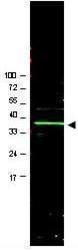
- Western blot using GeneTex's Affinity Purified anti-Bmi1 antibody shows detection of a band ~37 kDa corresponding to human Bmi1 (arrowhead). Approximately 20 µg of a U2OS whole cell lysate (bone osteosarcoma) was separated by 4-20% SDS-PAGE and transferred onto nitrocellulose. After blocking in PBS containing 5% nonfat dry milk, the membrane was probed overnight at 4° C with the primary antibody diluted to 1:1,000 in PBS containing 1% nonfat dry milk. The membrane was washed and reacted with a 1:20,000 dilution of IRDye800 conjugated rabbit anti-Goat IgG [H&L] for 45 min at room temperature. IRDye800 fluorescence image was captured using the Odyssey® Infrared Imaging System developed by LI-COR. IRDye is a trademark of LI-COR, Inc. Other detection systems will yield similar results.
- Validation comment
- WB

- Submitted by
- GeneTex (provider)
- Main image

- Experimental details
- Western blot using GeneTex's Affinity Purified anti-Bmi1 antibody shows detection of a band ~37 kDa corresponding to human Bmi1 (arrowhead). Approximately 20 ?g of a U2OS whole cell lysate was separated by 4-20% SDS-PAGE and transferred onto nitrocellulose. After blocking in PBS containing 5% nonfat dry milk, the membrane was probed overnight at 4¢X C with the primary antibody diluted to 1:1,000 in PBS containing 1% nonfat dry milk. The membrane was washed and reacted with a 1:20,000 dilution of IRDye800 conjugated rabbit anti-Goat IgG [H&L] for 45 min at room temperature. IRDye800 fluorescence image was captured using the Odyssey? Infrared Imaging System developed by LI-COR. IRDye is a trademark of LI-COR, Inc. Other detection systems will yield similar results.

Supportive validation
- Submitted by
- GeneTex (provider)
- Main image

- Experimental details
- Immunofluorescence using affinity purified goat anti Bmi1(GTX45791) shows nuclear staining (green) of methanol fixed (100%, 5 min) HepG2 cells. The cells were blocked and permeabilized in 1%BSA / 10% normal donkey serum / 0.3M glycine in 0.1% PBS-Tween for 1h prior to incubation with the primary antibody (1:200 dilution) overnight at +4¢XC and detected with a 488nm fluorescent dye conjugated secondary Ab. Cell nuclei are stained with DAPI (blue) and plasma membranes are stained with WGA (red).
