 Explore
 Validate
 Learn
Learn Western blot
Antibody data
- Antibody Data
- Antigen structure
- References [0]
- Comments [0]
- Validations
- Western blot [1]
- Immunocytochemistry [1]
Submit
Validation data
Reference
Comment
Report error
- Product number
- TA319184 - Provider product page

- Provider
- OriGene
- Product name
- Goat polyclonal anti-BMI1 antibody
- Antibody type
- Polyclonal
- Description
- Goat polyclonal anti-BMI1 antibody
- Host
- Goat
- Conjugate
- Unconjugated
- Epitope
- BMI1
- Antibody clone number
- NULL
- Vial size
- 100 µg
- Concentration
- 0.91 mg/mL
No comments: Submit comment
Supportive validation
- Submitted by
- OriGene (provider)
- Main image
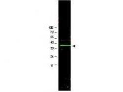
- Experimental details
- WB using Anti-Bmi1 antibody shows detection of a band ~37 kDa corresponding to human Bmi1 (arrowhead).Approximately 20 ug of a U2OS whole cell lysate (bone osteosarcoma) was separated by 4-20% SDS-PAGE and transferred onto nitrocellulose.?The primary antibody diluted to 1:1,000 in PBS containing 1% nonfat dry milk.? The membrane was washed and reacted with a 1:20,000 dilution of IRDye?800 conjugated Rb-a-Goat IgG [H&L] MX for 45 min at room temperature.
- Validation comment
- WB

Supportive validation
- Submitted by
- OriGene (provider)
- Main image

- Experimental details
- Immunofluorescence using affinity purified goat anti Bmi1 shows nuclear staining (green) of methanol fixed (100%, 5 min) HepG2 cells. The cells were blocked and permeabilized in 1%BSA / 10% normal donkey serum / 0.3M glycine in 0.1% PBS-Tween for 1h prior to incubation with the primary antibody (1:200 dilution) overnight at +4°C and detected with a 488nm fluorescent dye conjugated secondary Ab. Cell nuclei are stained with DAPI (blue) and plasma membranes are stained with WGA (red).
- Validation comment
- IF
