 Explore
 Validate
 Learn
Learn Western blot
 ELISA
ELISAAntibody data
- Antibody Data
- Antigen structure
- References [0]
- Comments [0]
- Validations
- Western blot [3]
- Immunocytochemistry [3]
- Immunohistochemistry [1]
- Flow cytometry [1]
Submit
Validation data
Reference
Comment
Report error
- Product number
- MA5-17053 - Provider product page

- Provider
- Invitrogen Antibodies
- Product name
- CDK5 Monoclonal Antibody (4E4)
- Antibody type
- Monoclonal
- Antigen
- Purifed from natural sources
- Description
- MA5-17053 targets CDK5 in indirect ELISA, FACS, ICC, IHC, IF and WB applications and shows reactivity with Human, Non-human primate, and Rat samples. The MA5-17053 immunogen is purified recombinant fragment of human CDK5 expressed in E. Coli. MA5-17053 detects CDK5 which has a predicted molecular weight of approximately 35kDa.
- Reactivity
- Human, Mouse, Rat
- Host
- Mouse
- Isotype
- IgG
- Antibody clone number
- 4E4
- Vial size
- 100 µL
- Concentration
- Conc. Not Determined
- Storage
- Store at 4°C short term. For long term storage, store at -20°C, avoiding freeze/thaw cycles.
No comments: Submit comment
Supportive validation
- Submitted by
- Invitrogen Antibodies (provider)
- Main image
- Experimental details
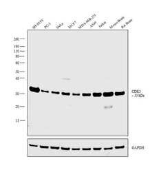
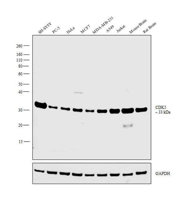
- Western blot analysis was performed on whole cell extracts (30 µg lysate) of SH-SY5Y (Lane 1), PC-3 (Lane 2), HeLa (Lane 3), MCF7 (Lane 4), MDA-MB-231 (Lane 5), A549 (Lane 6), Jurkat (Lane 7), tissue extracts of Mouse Brain (Lane 8) and Rat Brain (Lane 9). The blot was probed with CDK5 Monoclonal Antibody (Product # MA5-17053, 1:500 dilution) and detected by chemiluminescence using Goat anti-Mouse IgG (H+L) Superclonal™ Secondary Antibody, HRP conjugate (Product # A28177, 0.25 µg/ml, 1:4000 dilution). A band at 33kDa corresponding to CDK5 was observed across the cell lines and tissues tested.
- Submitted by
- Invitrogen Antibodies (provider)
- Main image

- Experimental details
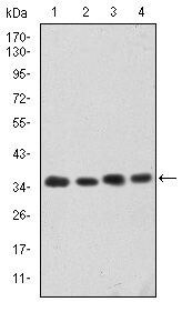
- Western blot analysis of CDK5 using CDK5 monoclonal antibody (Product # MA5-17053) in HeLa (1), K562 (2), PC-12 (3) and COS-7 (4) cell lysate.
- Submitted by
- Invitrogen Antibodies (provider)
- Main image
- Experimental details
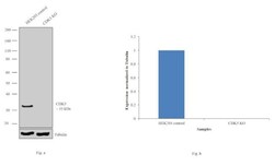
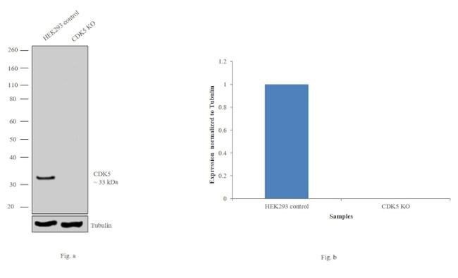
- Western blot analysis of CDK5 was performed by loading 30 ug of HEK293 control (Lane 1), HEK293- CDK5 knockout (Lane 2) edigene lysates. The blot was probed with CDK5 Monoclonal Antibody (4E4) (Product # MA5-17053, 1:1000 dilution) and Goat anti-Mouse IgG (H+L) Superclonal™ Secondary Antibody, HRP conjugate (Product # A28177, 0.25 µg/ml, 1:4000 dilution). Loss of signal upon CRISPR mediated knockout (KO) confirms that antibody is specific to CDK5.
Supportive validation
- Submitted by
- Invitrogen Antibodies (provider)
- Main image
- Experimental details
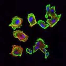
- Immunofluorescence analysis of GC7901 cells using CDK5 monoclonal antibody (Product # MA5-17053) (Green). Blue: DRAQ5 fluorescent DNA dye. Red: actin filaments have been labeled with phalloidin.

- Submitted by
- Invitrogen Antibodies (provider)
- Main image

- Experimental details
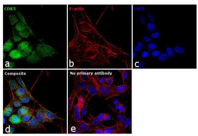
- Immunofluorescence analysis of CDK5 was performed using 70% confluent log phase SH-SY5Y cells. The cells were fixed with 4% paraformaldehyde for 10 minutes, permeabilized with 0.1% Triton™ X-100 for 15 minutes, and blocked with 1% BSA for 1 hour at room temperature. The cells were labeled with CDK5 Monoclonal Antibody (4E4) (Product # MA5-17053) at 1:100 dilution in 0.1% BSA, incubated at 4 degree Celsius overnight and then labeled with Goat anti-Mouse IgG (H+L) Superclonal™ Secondary Antibody, Alexa Fluor® 488 conjugate (Product # A28175) at a dilution of 1:2000 for 45 minutes at room temperature (Panel a: green). Nuclei (Panel b: blue) were stained with ProLong™ Diamond Antifade Mountant with DAPI (Product # P36962). F-actin (Panel c: red) was stained with Rhodamine Phalloidin (Product # R415). Panel d represents the merged image showing predominant nuclear localization. The images were captured at 60X magnification.
- Submitted by
- Invitrogen Antibodies (provider)
- Main image

- Experimental details
- Immunofluorescence analysis of GC7901 cells using CDK5 monoclonal antibody (Product # MA5-17053) (Green). Blue: DRAQ5 fluorescent DNA dye. Red: actin filaments have been labeled with phalloidin.

Supportive validation
- Submitted by
- Invitrogen Antibodies (provider)
- Main image

- Experimental details
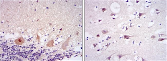
- Immunohistochemical analysis of paraffin-embedded cerebellum tissues (left) and brain tissues (right) using CDK5 monoclonal antibody (Product # MA5-17053) followed with DAB staining.
Supportive validation
- Submitted by
- Invitrogen Antibodies (provider)
- Main image

- Experimental details
- Flow cytometric analysis of K562 cells using CDK5 monoclonal antibody (Product # MA5-17053) (green) and negative control (purple).
