Explore
 Validate
 Learn
Learn Western blot
Antibody data
- Antibody Data
- Antigen structure
- References [0]
- Comments [0]
- Validations
- Western blot [1]
- Immunocytochemistry [1]
- Immunohistochemistry [1]
- Flow cytometry [1]
Submit
Validation data
Reference
Comment
Report error
- Product number
- Ab095769 - Provider product page

- Provider
- Aladdin Scientific
- Product name
- CDK5 Mouse mAb
- Antibody type
- Monoclonal
- Description
- Mouse anti human CDK5 Antibody, Monoclonal (1552CT262.105.8), could be used for WB, IHC, Flow, ICC, IF and so on.ApplicationsWB: 1/2000IHC: 1/100-1/500IF/ICC: 1/25Flow_1/25FunctionProbably involved in the control of the cell cycle. Interacts with D1 and D3-type G1 cyclins. Can phosphorylate histone H1, tau, MAP2 and NF-H and NF-M. Also interacts with p35 which activates the kinase.
- Reactivity
- Human, Mouse, Rat
- Host
- Mouse
- Conjugate
- Unconjugated
- Antigen sequence
AA 1-292- Antibody clone number
- 1552CT262.105.8
- Vial size
- 100_l,10_l,1ml,50_l
- Concentration
- 0,5 mg/ml
- Storage
- Store at 4¡C short term (1-2 weeks). Store at -20¡C long term (24 months). Upon delivery aliquot. Avoid freeze/thaw cycle.
No comments: Submit comment
Supportive validation
- Submitted by
- Aladdin Scientific (provider)
- Main image
- Experimental details
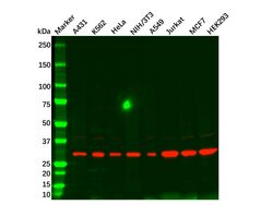
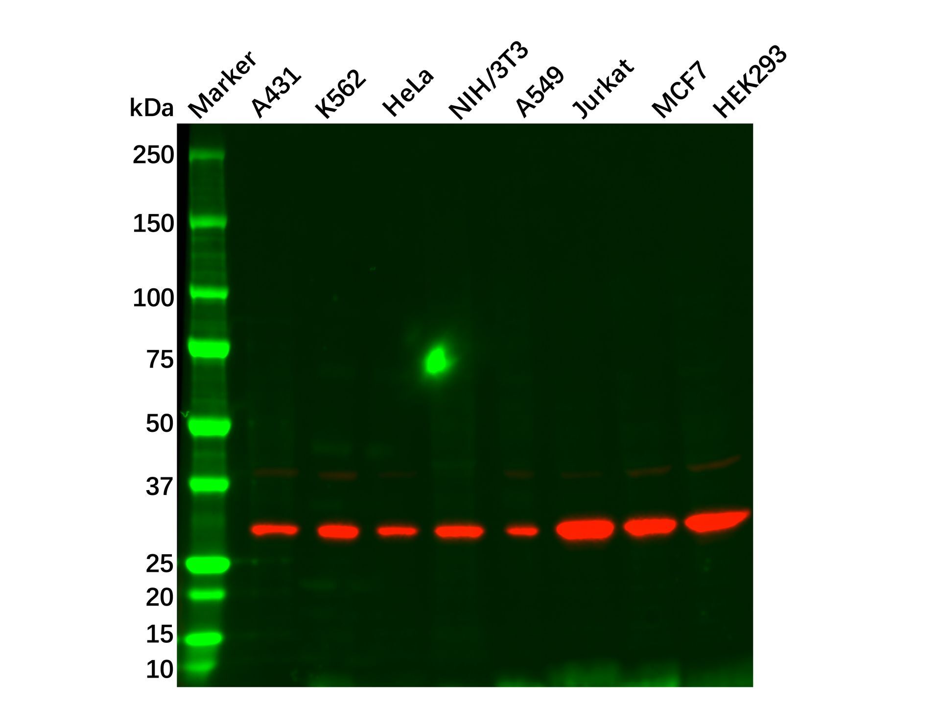
- CDK5 Mouse mAb (Ab095769) - Western Blot All lanes: CDK5 Mouse mAb (Ab095769) at 1/1000 dilution Samples: Lysates at 20 µg per lane Secondary: Goat Anti-Mouse IgG H&L (HRP) (Ab138040) at 1/40000 dilution Predicted band size: 33 kDa Observed band size: 30 kDa Exposure time: 11.9 s
Supportive validation
- Submitted by
- Aladdin Scientific (provider)
- Main image
- Experimental details
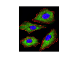
- CDK5 Mouse mAb (Ab095769) - ICC/IF Immunofluorescent analysis of 4% paraformaldehyde-fixed, 0.1% Triton X-100 permeabilized A549 (human lung adenocarcinoma epithelial cell line) cells labeling CDK5 with CDK5 Mouse mAb (Ab095769) at 1/25 dilution, followed by Dylight® 488-conjugated goat anti-mouse IgG secondary antibody at 1/200 dilution (green). Immunofluorescence image showing cytoplasm staining on A549 cell line. Cytoplasmic actin is detected with Dylight® 554 Phalloidin at 1/100 dilution (red). The nuclear counter stain is DAPI (blue).

Supportive validation
- Submitted by
- Aladdin Scientific (provider)
- Main image
- Experimental details
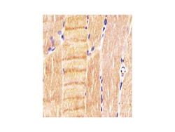
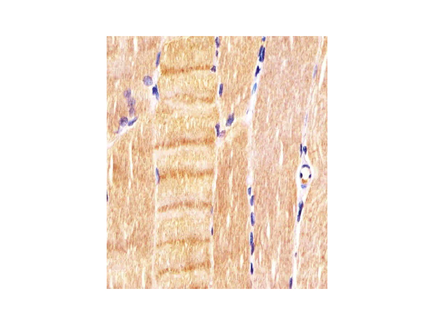
- CDK5 Mouse mAb (Ab095769) - IHC CDK5 Mouse mAb (Ab095769) staining CDK5 in human skeletal muscle sections by Immunohistochemistry (paraformaldehyde - fixed, paraffin - embedded sections). Tissue was fixed with formaldehyde and blocked with 3% BSA for 0.5 hour at room temperature; antigen retrieval was by heat mediation with a citrate buffer (pH 6). Samples were incubated with primary antibody (1/25) for 1 hours at 37°C. A undiluted biotinylated goat polyvalent antibody was used as the secondary antibody.
Supportive validation
- Submitted by
- Aladdin Scientific (provider)
- Main image
- Experimental details
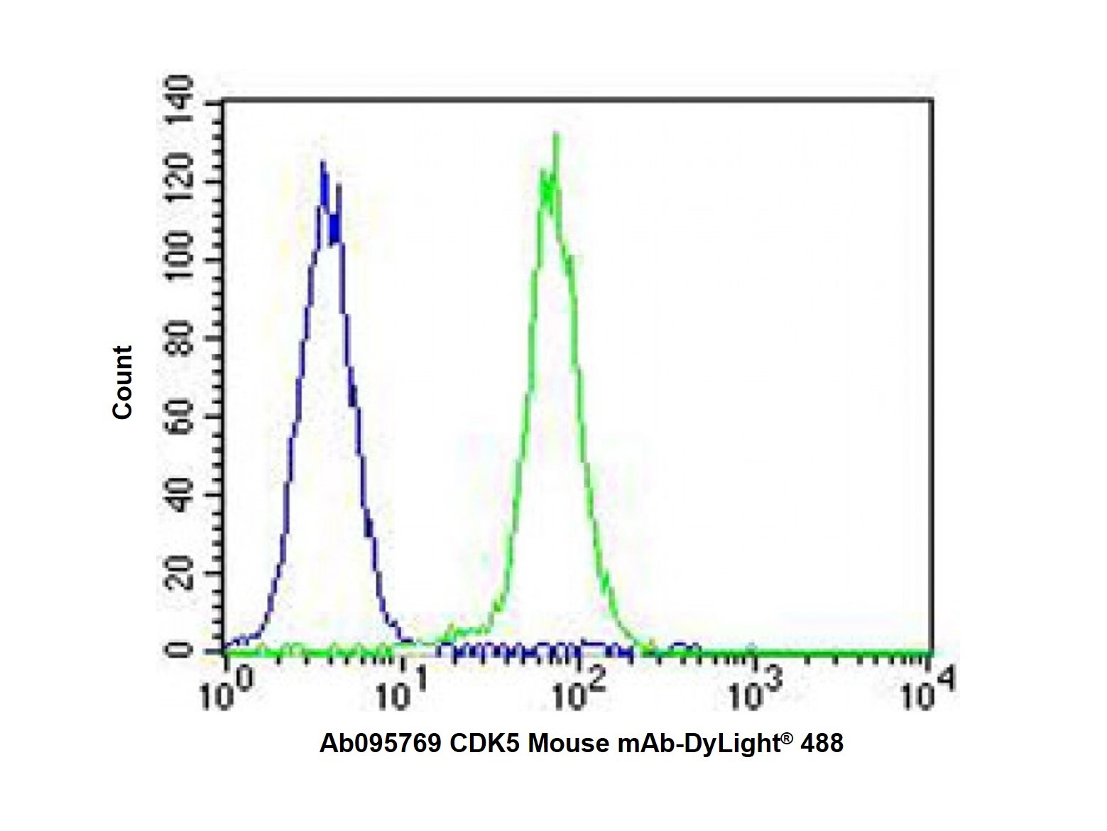
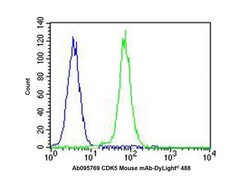
- CDK5 Mouse mAb (Ab095769) - Flow Cytometry Overlay histogram showing K562 cells stained with CDK5 Mouse mAb (Ab095769) (green line). The cells were fixed with 2% paraformaldehyde (10 min) and then permeabilized with 90% methanol for 10 min. The cells were then incubated in 2% bovine serum albumin to block non-specific protein-protein interactions followed by CDK5 Mouse mAb (Ab095769) (1/25 dilution) for 60 min at 37ºC. The secondary antibody was used Goat-Anti-Mouse IgG, DyLight® 488 Conjugated Highly Cross-Adsorbed at 1/400 dilution for 40 min at 37ºC. Isotype control antibody (blue line) was mouse IgG1 used under the same conditions. Acquisition of >10000 events was performed.