 Explore
 Validate
 Learn
Learn Western blot
 Immunocytochemistry
Antibody data
- Antibody Data
- Antigen structure
- References [0]
- Comments [0]
- Validations
- Immunocytochemistry [4]
Submit
Validation data
Reference
Comment
Report error
- Product number
- PA5-17218 - Provider product page

- Provider
- Invitrogen Antibodies
- Product name
- Anti-Atg12
- Antibody type
- Polyclonal
- Antigen
- Synthetic peptide corresponding to residues near the amino terminus of mouse Atg12.
- Host
- Rabbit
- Vial size
- 100 ul
- Storage
- -20°C
No comments: Submit comment
Supportive validation
- Submitted by
- Invitrogen Antibodies (provider)
- Main image
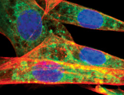
- Experimental details
- Immunofluorescent analysis of Atg12 in NIH/3T3 cells using an Atg12 polyclonal antibody (Product # PA5-17218) (green). Actin filaments are labeled with a fluorescent red phalloidin. DNA is labeled using a fluorescent blue dye.
- Submitted by
- Invitrogen Antibodies (provider)
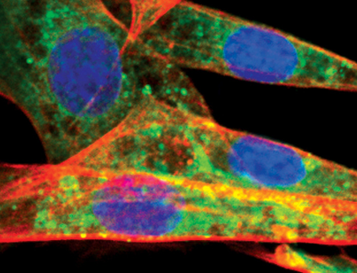
- Main image
- Experimental details
- Immunofluorescent analysis of Atg12 in NIH/3T3 cells, chloroquine-treated, using an Atg12 polyclonal antibody (Product # PA5-17218) (green). Actin filaments are labeled with a fluorescent red phalloidin. DNA is labeled using a fluorescent blue dye.

- Submitted by
- Invitrogen Antibodies (provider)
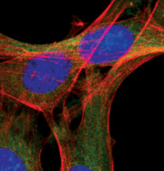
- Main image

- Experimental details
- Immunofluorescent analysis of Atg12 in NIH/3T3 cells, chloroquine-treated, using an Atg12 polyclonal antibody (Product # PA5-17218) (green). Actin filaments are labeled with a fluorescent red phalloidin. DNA is labeled using a fluorescent blue dye.
- Submitted by
- Invitrogen Antibodies (provider)
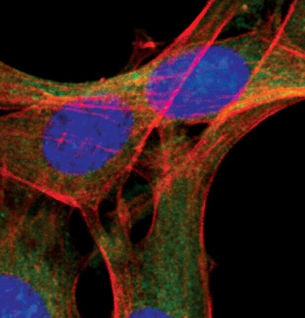
- Main image
- Experimental details
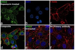
- Immunofluorescence analysis of ATG12 was performed using 70% confluent log phase NIH/3T3 cells treated with 50uM of Rapamycin for 16 hours. The cells were fixed with 4% paraformaldehyde for 10 minutes, permeabilized with 0.1% Triton™ X-100 for 15 minutes, and blocked with 2% BSA for 45 minutes at room temperature. The cells were labeled with ATG12 Polyclonal Antibody (Product # PA5-17218) at 1:100 in 0.1% BSA, incubated at 4 degree celsius overnight and then labeled with Donkey anti-Rabbit IgG (H+L) Highly Cross-Adsorbed Secondary Antibody, Alexa Fluor Plus 488 (Product # A32790), (1:2000), for 45 minutes at room temperature (Panel a: Green). Nuclei (Panel b:Blue) were stained with ProLong™ Diamond Antifade Mountant with DAPI (Product # P36962). F-actin (Panel c: Red) was stained with Rhodamine Phalloidin (Product # R415, 1:300). Panel d represents the merged image showing cytoplasmic localization. Panel e represents untreated cells with weak cytoplasmic signal Panel f represents control cells with no primary antibody to assess background. The images were captured at 60X magnification.
