Explore
 Validate
 Learn
Learn Western blot
 Immunocytochemistry
Antibody data
- Antibody Data
- Antigen structure
- References [0]
- Comments [0]
- Validations
- Immunocytochemistry [4]
- Immunohistochemistry [2]
Submit
Validation data
Reference
Comment
Report error
- Product number
- PA5-38036 - Provider product page

- Provider
- Invitrogen Antibodies
- Product name
- NOX3 Polyclonal Antibody
- Antibody type
- Polyclonal
- Antigen
- Synthetic peptide
- Description
- PA5-38036 detects Nox3 in mouse bone marrow and humanG-CSF mobilized CD34+ cells in immunofluorescence analysis. A recommended positive control is 293 cell lysate. NOX3 is not expected to cross-react with other NOX proteins.
- Reactivity
- Human, Mouse
- Host
- Rabbit
- Isotype
- IgG
- Vial size
- 100 μg
- Concentration
- 1 mg/mL
- Storage
- Maintain refrigerated at 2-8°C for up to 3 months. For long term storage store at -20°C
No comments: Submit comment
Supportive validation
- Submitted by
- Invitrogen Antibodies (provider)
- Main image
- Experimental details
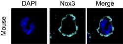
- Immunofluorescent analysis of Nox3 (Violet) in mouse bone marrow cells (Up). The cells were fixed with 2% paraformaldehyde in PBS for 30 minutes at RT, permeabilized with 0.5% Triton X-100 in PBS for 30 minutes at RT, and blocked with blocking buffer for 4 hours in cold room. Cells were stained with rabbit polyclonal anti-Nox3 antibody (Product # PA5-38036) at a dilution of 1:200 in blocking buffer overnight in cold room, wash 5 times with 1 xPBS, incubated with goat anti-rabbit IgG antibody conjugated with Cy5 (Product # A10523) at a dilution of 1:1000 for 1 hours at RT (panel b: Violet) and wash 5 times with 1xPBS before overlaid with cover slip. Nuclei (panel a: blue) were stained with DAPI (Product # D1306) during the blocking step. Panels a and b are a merged image and magnified squire area, respectively. Images were taken on a Nikon A1 confocal microscopy at 60x Objective lens with oil used. Data courtesy of Dr. Zhi Wen from the University of Wisconsin, Madison.

- Submitted by
- Invitrogen Antibodies (provider)
- Main image

- Experimental details
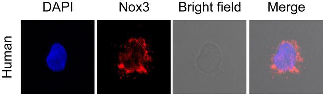
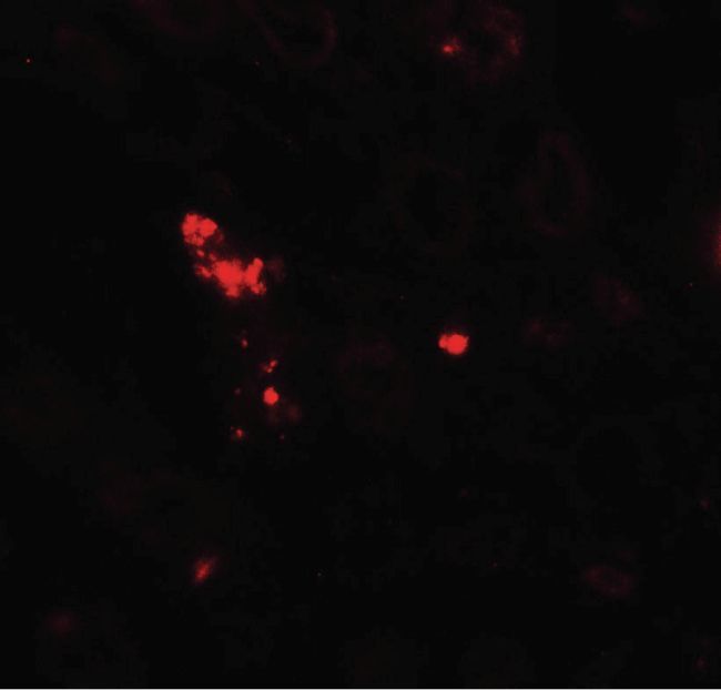
- Immunofluorescent analysis of Nox3 (Red) in human G-CSF mobilized CD34+ cells. The cells were fixed with 2% paraformaldehyde in PBS for 30 minutes at RT, permeabilized with 0.5% Triton X-100 in PBS for 30 minutes at RT, and blocked with blocking buffer for 4 hours in cold room. Cells were stained with rabbit polyclonal anti-Nox3 antibody (Product # PA5-38036) at a dilution of 1:200 in blocking buffer overnight in cold room, wash 5 times with 1 xPBS, incubated with goat anti-rabbit IgG antibody conjugated with Dylight 594 (Product # 35560) at a dilution of 1:1000 for 1 hours at RT (panel b: red) and wash 5 times with 1xPBS before overlaid with cover slip. Nuclei (panel a: blue) were stained with DAPI (Product # D1306) during the blocking step. Panel c was the bright field. Panels a, b and c are a merged image and magnified squire area, respectively. Images were taken on a Nikon A1 confocal microscopy at 60x Objective lens with oil used. Data courtesy of Dr. Zhi Wen from the University of Wisconsin, Madison.

- Submitted by
- Invitrogen Antibodies (provider)
- Main image

- Experimental details
- Immunofluorescent analysis of Nox3 (Red) in human G-CSF mobilized CD34+ cells. The cells were fixed with 2% paraformaldehyde in PBS for 30 minutes at RT, permeabilized with 0.5% Triton X-100 in PBS for 30 minutes at RT, and blocked with blocking buffer for 4 hours in cold room. Cells were stained with rabbit polyclonal anti-Nox3 antibody (Product # PA5-38036) at a dilution of 1:200 in blocking buffer overnight in cold room, wash 5 times with 1 xPBS, incubated with goat anti-rabbit IgG antibody conjugated with Dylight 594 (Product # 35560) at a dilution of 1:1000 for 1 hours at RT (panel b: red) and wash 5 times with 1xPBS before overlaid with cover slip. Nuclei (panel a: blue) were stained with DAPI (Product # D1306) during the blocking step. Panel c was the bright field. Panels a, b and c are a merged image and magnified squire area, respectively. Images were taken on a Nikon A1 confocal microscopy at 60x Objective lens with oil used. Data courtesy of Dr. Zhi Wen from the University of Wisconsin, Madison.
- Submitted by
- Invitrogen Antibodies (provider)
- Main image

- Experimental details
- Immunofluorescent analysis of Nox3 (Violet) in mouse bone marrow cells (Up). The cells were fixed with 2% paraformaldehyde in PBS for 30 minutes at RT, permeabilized with 0.5% Triton X-100 in PBS for 30 minutes at RT, and blocked with blocking buffer for 4 hours in cold room. Cells were stained with rabbit polyclonal anti-Nox3 antibody (Product # PA5-38036) at a dilution of 1:200 in blocking buffer overnight in cold room, wash 5 times with 1 xPBS, incubated with goat anti-rabbit IgG antibody conjugated with Cy5 (Product # A10523) at a dilution of 1:1000 for 1 hours at RT (panel b: Violet) and wash 5 times with 1xPBS before overlaid with cover slip. Nuclei (panel a: blue) were stained with DAPI (Product # D1306) during the blocking step. Panels a and b are a merged image and magnified squire area, respectively. Images were taken on a Nikon A1 confocal microscopy at 60x Objective lens with oil used. Data courtesy of Dr. Zhi Wen from the University of Wisconsin, Madison.

Supportive validation
- Submitted by
- Invitrogen Antibodies (provider)
- Main image
- Experimental details
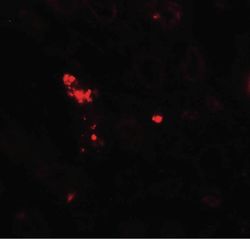
- Immunofluorescence of NOX3 in human kidney tissue with NOX3 Polyclonal Antibody (Product # PA5-38036) at 20 µg/mL.
- Submitted by
- Invitrogen Antibodies (provider)
- Main image
- Experimental details
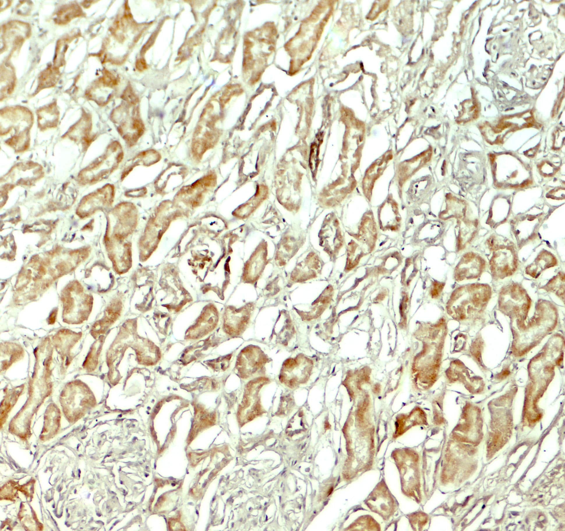
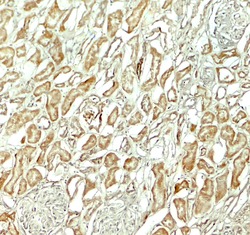
- Immunohistochemistry of NOX3 in human kidney tissue with NOX3 Polyclonal Antibody (Product # PA5-38036) at 5 µg/mL.