Explore
 Validate
 Learn
Learn Western blot
Antibody data
- Antibody Data
- Antigen structure
- References [1]
- Comments [0]
- Validations
- Western blot [1]
- Flow cytometry [2]
- Other assay [1]
Submit
Validation data
Reference
Comment
Report error
- Product number
- PA5-25614 - Provider product page

- Provider
- Invitrogen Antibodies
- Product name
- NTCP Polyclonal Antibody
- Antibody type
- Polyclonal
- Antigen
- Synthetic peptide
- Reactivity
- Human
- Host
- Rabbit
- Isotype
- IgG
- Vial size
- 200 μL
- Concentration
- 0.5 mg/mL
- Storage
- Store at 4°C short term. For long term storage, store at -20°C, avoiding freeze/thaw cycles.
Submitted references Robust in vitro assay for analyzing the neutralization activity of serum specimens against hepatitis B virus.
Zhang YL, Gao Y, Cao JL, Zhao JH, Zhang TY, Yang CL, Xiong HL, Wang YB, Ou SH, Cheng T, Chen CR, Yuan Q, Xia NS
Emerging microbes & infections 2019;8(1):724-733
Emerging microbes & infections 2019;8(1):724-733
No comments: Submit comment
Supportive validation
- Submitted by
- Invitrogen Antibodies (provider)
- Main image
- Experimental details
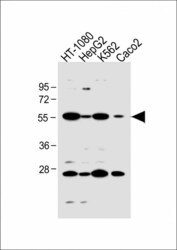
- Western blot analysis of NTCP in various lysates. Samples were incubated with NTCP polyclonal antibody (Product # PA5-25614) using a dilution of 1:1,000 followed by Goat Anti-Rabbit IgG, (H+L), Peroxidase conjugated at a dilution of 1:10,000. Lysates/proteins: 20 µg per lane. Lane 1: HT-1080 whole cell lysate; Lane 2: HepG2 whole cell lysate; Lane 3: K562 whole cell lysate; Lane 4: Caco2 whole cell lysate. Predicted band size: 38 kDa. Blocking/Dilution buffer: 5% NFDM/TBST.

Supportive validation
- Submitted by
- Invitrogen Antibodies (provider)
- Main image
- Experimental details
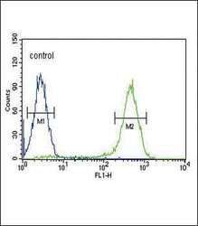
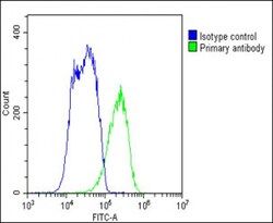
- Flow cytometry analysis of K562 cells using a SLC10A1 polyclonal antibody (Product # PA5-25614) (right) compared to a negative control cell (left) at a dilution of 1:10-50, followed by a FITC-conjugated goat anti-rabbit antibody
- Submitted by
- Invitrogen Antibodies (provider)
- Main image
- Experimental details
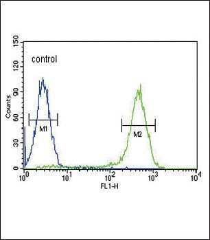
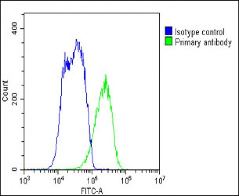
- Flow cytometry of (overlay histogram) of NTCP in HepG2 cells (green line). Samples were incubated with NTCP polyclonal antibody (Product # PA5-25614) using a dilution of 1:25 dilution for 60 min at 37°C followed by Goat-Anti-Rabbit IgG, DyLight® 488 Conjugated Highly Cross-Adsorbed at 1:200 dilution for 40 min at 37°C. The cells were fixed with 2% paraformaldehyde 10 min. The cells were then incubated in 2% bovine serum albumin to block non-specific protein-protein interactions followed by the primary antibody. Isotype control antibody (blue line) was rabbit IgG1 (1 μg/1x10^6 cells) used under the same conditions. Acquisition of >10, 000 events was performed.
Supportive validation
- Submitted by
- Invitrogen Antibodies (provider)
- Main image

- Experimental details
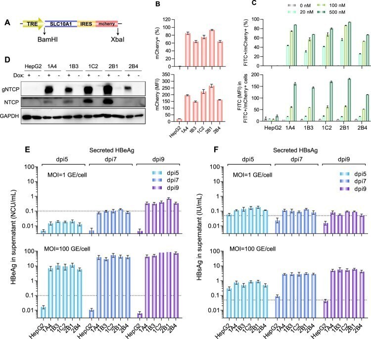
- Figure 1. Establishment and characterization of NTCP-reconstituted HepG2 cells for in vitro HBV infection. (A) A schematic diagram depicting the dox-inducible NTCP expression cassette. The NTCP-IRES-mCherry cassette was cloned into the pLenti-CMVTRE3G-eGFP (Addgene 27570) vector between the BamHI and XbaI restriction sites. (B) Flow cytometric analyses of the dox-induced mCherry expression of the different HepG2-TetOn-NTCP cell lines. The upper panel showed the percentage of mCherry+ cell population, and the lower panel showed the mean fluorescence intensity (MFI) of mCherry+ cell population of various cell lines. (C) Flow cytometric analyses of the dox-induced PreS1-peptide (FITC conjugated) binding capability of the different HepG2-TetOn-NTCP cell lines. The upper panel presented the dose-dependent comparison on the FITC/mCherry double positive percentage of different cell lines, and the lower panel presented the FITC MFI of FITC+/mCherry+ cell populations of various cell lines when binding with myr-PreS1-FITC peptide at different dosage (500, 100 and 20 nM). (D) Western blots for NTCP expression in the different HepG2-TetOn-NTCP cell lines with or without dox induction. The NTCP was detected by a commercial rabbit polyclonal antibody against NTCP (Thermo Fisher Scientific, PA525614). (E, F) Detections of HBeAg (E) and HBsAg (F) in culture supernatants of different HepG2-TetOn-NTCP cell lines following infection with HBV at 1 GE/cell (upper panel) or 100 GE/cell (lower pa