Explore
 Validate
 Learn
LearnNBP1-89954
antibody from Novus Biologicals
Targeting: DMD
BMD, DXS142, DXS164, DXS206, DXS230, DXS239, DXS268, DXS269, DXS270, DXS272, MRX85
 Immunohistochemistry
Antibody data
- Antibody Data
- Antigen structure
- References [0]
- Comments [0]
- Validations
- Immunohistochemistry [5]
Submit
Validation data
Reference
Comment
Report error
- Product number
- NBP1-89954 - Provider product page

- Provider
- Novus Biologicals
- Proper citation
- Novus Cat#NBP1-89954, RRID:AB_11030305
- Product name
- Rabbit Polyclonal Dystrophin Antibody
- Antibody type
- Polyclonal
- Description
- Immunogen affinity purified. Specificity of human Dystrophin antibody verified on a Protein Array containing target protein plus 383 other non-specific proteins.
- Reactivity
- Human
- Host
- Rabbit
- Isotype
- IgG
- Vial size
- 0.1 ml
- Storage
- Store at 4C short term. Aliquot and store at -20C long term. Avoid freeze-thaw cycles.
No comments: Submit comment
Supportive validation
- Submitted by
- Novus Biologicals (provider)
- Main image

- Experimental details
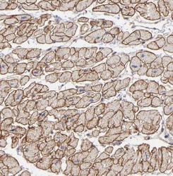
- Immunohistochemistry-Paraffin: Dystrophin Antibody [NBP1-89954] - Staining of human heart muscle shows strong cytoplasmic and membranous positivity in myocytes.
- Submitted by
- Novus Biologicals (provider)
- Main image
- Experimental details
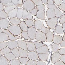
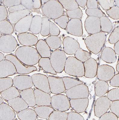
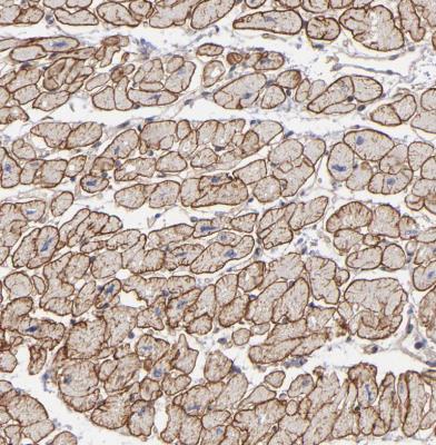
- Immunohistochemistry-Paraffin: Dystrophin Antibody [NBP1-89954] - Staining of human skeletal muscle shows moderate membranous positivity in myocytes.
- Submitted by
- Novus Biologicals (provider)
- Main image
- Experimental details
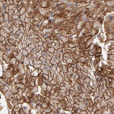
- Immunohistochemistry-Paraffin: Dystrophin Antibody [NBP1-89954] - Staining of human heart muscle shows strong membranous positivity in cardiomyocytes.
- Submitted by
- Novus Biologicals (provider)
- Main image
- Experimental details
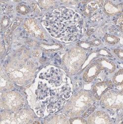
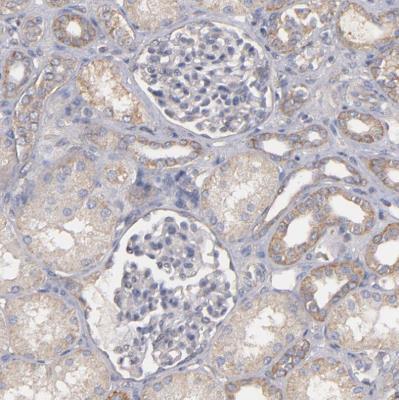
- Immunohistochemistry-Paraffin: Dystrophin Antibody [NBP1-89954] - Staining of human kidney shows no positivity in cells in glomeruli.
- Submitted by
- Novus Biologicals (provider)
- Main image
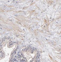
- Experimental details
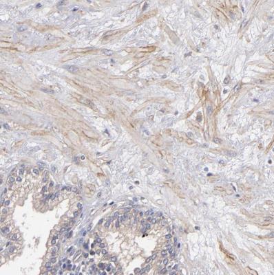
- Immunohistochemistry-Paraffin: Dystrophin Antibody [NBP1-89954] - Staining of human prostate shows weak membranous positivity in smooth muscle cells.