Explore
 Validate
 Learn
Learn Western blot
 Immunocytochemistry
Antibody data
- Antibody Data
- Antigen structure
- References [5]
- Comments [0]
- Validations
- Western blot [2]
- Immunohistochemistry [2]
Submit
Validation data
Reference
Comment
Report error
- Product number
- NBP2-24779 - Provider product page

- Provider
- Novus Biologicals
- Product name
- Rabbit Polyclonal CXCR7/RDC-1 Antibody
- Antibody type
- Polyclonal
- Description
- Peptide affinity purified.
- Reactivity
- Human, Mouse, Rat, Sheep
- Host
- Rabbit
- Isotype
- IgG
- Vial size
- 0.1 mg
- Concentration
- 1.0 mg/ml
- Storage
- Store at 4C short term. Aliquot and store at -20C long term. Avoid freeze-thaw cycles.
Submitted references Inhibition of chemokine (C-X-C motif) receptor four (CXCR4) at the fetal-maternal interface during early gestation in sheep: alterations in expression of chemokines, angiogenic factors and their receptors.
The chemokine receptor CXCR7 is highly expressed in human glioma cells and mediates antiapoptotic effects.
CXCR7 is an active component of SDF-1 signalling in astrocytes and Schwann cells.
CXCR4 but not CXCR7 is mainly implicated in ocular leukocyte trafficking during ovalbumin-induced acute uveitis.
CXCL12 induction of inducible nitric oxide synthase in human CD8 T cells.
Quinn KE, Prosser SZ, Kane KK, Ashley RL
Journal of animal science 2017 Mar;95(3):1144-11153
Journal of animal science 2017 Mar;95(3):1144-11153
The chemokine receptor CXCR7 is highly expressed in human glioma cells and mediates antiapoptotic effects.
Hattermann K, Held-Feindt J, Lucius R, Müerköster SS, Penfold ME, Schall TJ, Mentlein R
Cancer research 2010 Apr 15;70(8):3299-308
Cancer research 2010 Apr 15;70(8):3299-308
CXCR7 is an active component of SDF-1 signalling in astrocytes and Schwann cells.
Odemis V, Boosmann K, Heinen A, Küry P, Engele J
Journal of cell science 2010 Apr 1;123(Pt 7):1081-8
Journal of cell science 2010 Apr 1;123(Pt 7):1081-8
CXCR4 but not CXCR7 is mainly implicated in ocular leukocyte trafficking during ovalbumin-induced acute uveitis.
Zhang Z, Zhong W, Hall MJ, Kurre P, Spencer D, Skinner A, O'Neill S, Xia Z, Rosenbaum JT
Experimental eye research 2009 Oct;89(4):522-31
Experimental eye research 2009 Oct;89(4):522-31
CXCL12 induction of inducible nitric oxide synthase in human CD8 T cells.
Choy JC, Yi T, Rao DA, Tellides G, Fox-Talbot K, Baldwin WM 3rd, Pober JS
The Journal of heart and lung transplantation : the official publication of the International Society for Heart Transplantation 2008 Dec;27(12):1333-9
The Journal of heart and lung transplantation : the official publication of the International Society for Heart Transplantation 2008 Dec;27(12):1333-9
No comments: Submit comment
Supportive validation
- Submitted by
- Novus Biologicals (provider)
- Main image

- Experimental details
- Simple Western: CXCR7/RDC-1 Antibody [NBP2-24779] - Lane view shows a specific band for CXCR7 in 0.5 mg/mL of Jurkat lysate. This experiment was performed under reducing conditions using the 12-230 kDa separation system.

- Submitted by
- Novus Biologicals (provider)
- Main image

- Experimental details
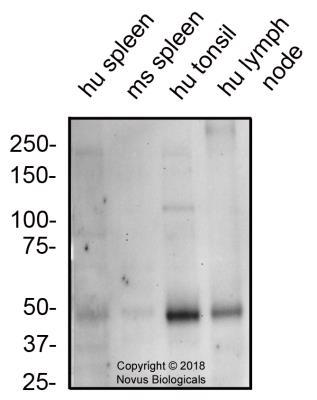
- Western Blot: CXCR7/RDC-1 Antibody [NBP2-24779] - Total protein from human spleen, tonsil, lymph node and mouse spleen was separated on a 7.5% gel by SDS-PAGE, transferred to PVDF membrane and blocked in 5% non-fat milk in TBST. The membrane was probed with 2.0 ug/mL anti-CXCR7 in 1% BSA in TBST and detected with an anti-rabbit HRP secondary antibody using chemiluminescence.
Supportive validation
- Submitted by
- Novus Biologicals (provider)
- Main image
- Experimental details
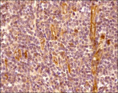
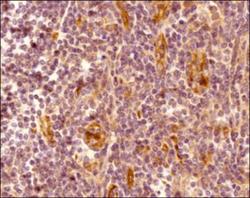
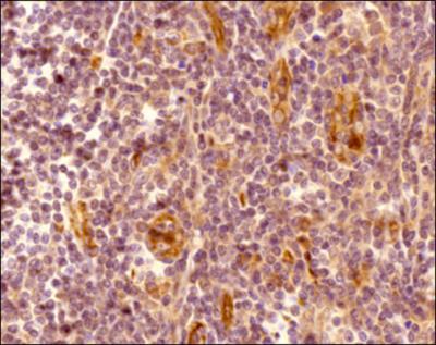
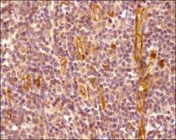
- Immunohistochemistry-Paraffin: CXCR7/RDC-1 Antibody [NBP2-24779] - Analysis of a FFPE tissue section of human tonsil using 5 ug/mL concentration of CXCR7/RDC-1 antibody. The staining was developed using HRP labeled anti-rabbit secondary antibody and DAB reagent, and nuclei of cells were counter-stained with hematoxylin. This CXCR7 antibody generated a specific membrane cytoplasmic staining in most of the cells, and the signal was highest/very intense in the endothelial cells/blood vessels.
- Submitted by
- Novus Biologicals (provider)
- Main image
- Experimental details
- Immunohistochemistry-Paraffin: CXCR7/RDC-1 Antibody [NBP2-24779] - Analysis of a FFPE tissue section of human tonsil using 5 ug/mL concentration of CXCR7/RDC-1 antibody. The staining was developed using HRP labeled anti-rabbit secondary antibody and DAB reagent, and nuclei of cells were counter-stained with hematoxylin. This CXCR7 antibody generated a specific membrane cytoplasmic staining in most of the cells, and the signal was highest/very intense in the endothelial cells/blood vessels.