 Explore
 Validate
 Learn
Learn Western blot
 Immunocytochemistry
Antibody data
- Antibody Data
- Antigen structure
- References [2]
- Comments [0]
- Validations
- Western blot [4]
- Immunohistochemistry [3]
Submit
Validation data
Reference
Comment
Report error
- Product number
- NBP1-31309 - Provider product page

- Provider
- Novus Biologicals
- Proper citation
- Novus Cat#NBP1-31309, RRID:AB_10003774
- Product name
- Rabbit Polyclonal CXCR7/RDC-1 Antibody
- Antibody type
- Polyclonal
- Description
- Immunogen affinity purified.
- Reactivity
- Human, Mouse
- Host
- Rabbit
- Isotype
- IgG
- Vial size
- 100 ul
- Storage
- Aliquot and store at -20C or -80C. Avoid freeze-thaw cycles.
Submitted references Correlation between CXCR4/CXCR7/CXCL12 chemokine axis expression and prognosis in lymph-node-positive lung cancer patients.
The IL-8-regulated chemokine receptor CXCR7 stimulates EGFR signaling to promote prostate cancer growth.
Katsura M, Shoji F, Okamoto T, Shimamatsu S, Hirai F, Toyokawa G, Morodomi Y, Tagawa T, Oda Y, Maehara Y
Cancer science 2018 Jan;109(1):154-165
Cancer science 2018 Jan;109(1):154-165
The IL-8-regulated chemokine receptor CXCR7 stimulates EGFR signaling to promote prostate cancer growth.
Singh RK, Lokeshwar BL
Cancer research 2011 May 1;71(9):3268-77
Cancer research 2011 May 1;71(9):3268-77
No comments: Submit comment
Supportive validation
- Submitted by
- Novus Biologicals (provider)
- Main image
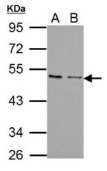
- Experimental details
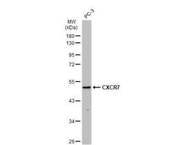
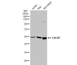
- Western Blot: CXCR7/RDC-1 Antibody [NBP1-31309] - Various whole cell extracts (30 ug) were separated by 10% SDS-PAGE, and the membrane was blotted with CXCR7 antibody [C1C2], Internal diluted at 1:1000. The HRP-conjugated anti-rabbit IgG antibody (NBP2-19301) was used to detect the primary antibody, and the signal was developed with Trident ECL plus-Enhanced.
- Submitted by
- Novus Biologicals (provider)
- Main image
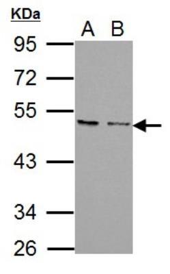
- Experimental details
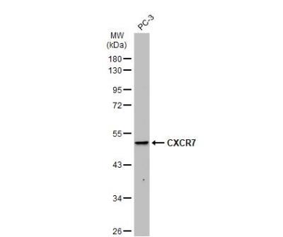
- Western Blot: CXCR7/RDC-1 Antibody [NBP1-31309] - Whole cell extract (30 ug) was separated by 10% SDS-PAGE, and the membrane was blotted with CXCR7 antibody [C1C2], Internal diluted at 1:500. The HRP-conjugated anti-rabbit IgG antibody (NBP2-19301) was used to detect the primary antibody.
- Submitted by
- Novus Biologicals (provider)
- Main image
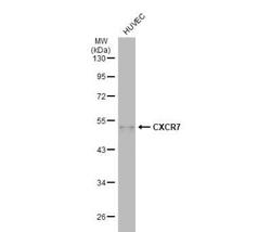
- Experimental details
- Western Blot: CXCR7/RDC-1 Antibody [NBP1-31309] - Whole cell extract (30 ug) was separated by 10% SDS-PAGE, and the membrane was blotted with CXCR7 antibody [C1C2], Internal diluted at 1:1000. The HRP-conjugated anti-rabbit IgG antibody (NBP2-19301was used to detect the primary antibody, and the signal was developed with Trident ECL plus-Enhanced.
- Submitted by
- Novus Biologicals (provider)
- Main image
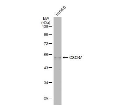
- Experimental details
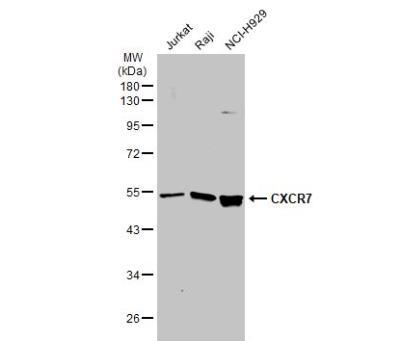
- Western Blot: CXCR7/RDC-1 Antibody [NBP1-31309] - Various whole cell extracts (30 ug) were separated by 10% SDS-PAGE, and the membrane was blotted with CXCR7 antibody [C1C2], Internal diluted at 1:1000. The HRP-conjugated anti-rabbit IgG antibody (NBP2-19301) was used to detect the primary antibody, and the signal was developed with Trident ECL plus-Enhanced.
Supportive validation
- Submitted by
- Novus Biologicals (provider)
- Main image
- Experimental details
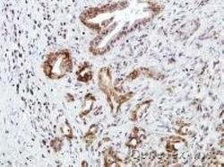
- Immunohistochemistry: CXCR7/RDC-1 Antibody [NBP1-31309] - Paraffin-embedded Human pancreatic tumor, using CXCR7 antibody at 1:100 dilution. 20X
- Submitted by
- Novus Biologicals (provider)
- Main image
- Experimental details
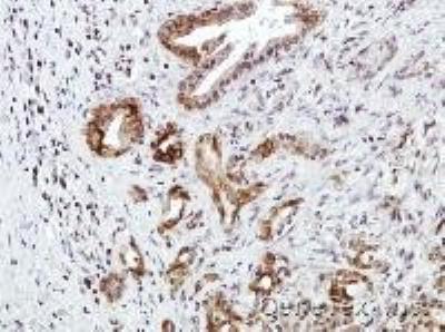
- Immunohistochemistry: CXCR7/RDC-1 Antibody [NBP1-31309] - Paraffin-embedded Human pancreatic tumor, using CXCR7antibody at 1:100 dilution. 40X

- Submitted by
- Novus Biologicals (provider)
- Main image
- Experimental details
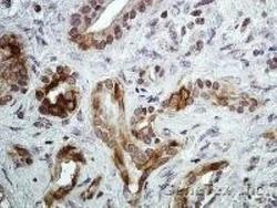
- Immunohistochemistry-Paraffin: CXCR7/RDC-1 Antibody [NBP1-31309] - Paraffin-embedded gastric CA, using antibody at 1:100 dilution.
