 Explore
 Validate
 Learn
Learn Western blot
Antibody data
- Antibody Data
- Antigen structure
- References [1]
- Comments [0]
- Validations
- Western blot [2]
- Immunocytochemistry [2]
- Immunohistochemistry [2]
Submit
Validation data
Reference
Comment
Report error
- Product number
- PA5-39887 - Provider product page

- Provider
- Invitrogen Antibodies
- Product name
- OTX2 Polyclonal Antibody
- Antibody type
- Polyclonal
- Antigen
- Recombinant full-length protein
- Description
- In direct ELISAs, approximately 10% cross-reactivity with recombinant human OTX1 is observed. Reconstitute at 0.2 mg/mL in sterile PBS.
- Reactivity
- Human
- Host
- Goat
- Isotype
- IgG
- Vial size
- 100 µg
- Concentration
- 0.2 mg/mL
- Storage
- -20°C or -80°C if preferred
Submitted references Constitutive activation of canonical Wnt signaling disrupts choroid plexus epithelial fate.
Parichha A, Suresh V, Chatterjee M, Kshirsagar A, Ben-Reuven L, Olender T, Taketo MM, Radosevic V, Bobic-Rasonja M, Trnski S, Holtzman MJ, Jovanov-Milosevic N, Reiner O, Tole S
Nature communications 2022 Feb 2;13(1):633
Nature communications 2022 Feb 2;13(1):633
No comments: Submit comment
Supportive validation
- Submitted by
- Invitrogen Antibodies (provider)
- Main image

- Experimental details
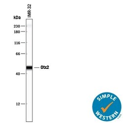
- Western blot analysis of OTX2 in IMR‚32 human neuroblastoma cell line. Samples were incubated in OTX2 polyclonal antibody (Product # PA5-39887) using a dilution of 1 µg/mL followed by a HRP-conjugated Anti-Goat IgG secondary antibody. A specific band was detected for Otx2 at approximately 37 kDa (as indicated). This experiment was conducted under reducing conditions.

- Submitted by
- Invitrogen Antibodies (provider)
- Main image
- Experimental details
- Western blot analysis of OTX2 in 0.2 mg/mL lysates of IMR‚32 human neuroblastoma cell line. Samples were incubated in OTX2 polyclonal antibody (Product # PA5-39887) using a dilution of 10 µg/mL followed by HRP-conjugated Anti-Goat IgG at a dilution of 0.0763888888888889. A specific band was detected for Otx2 at approximately 48 kDa (as indicated). This experiment was conducted under reducing conditions and using the 12-230 kDa separation system.

Supportive validation
- Submitted by
- Invitrogen Antibodies (provider)
- Main image

- Experimental details
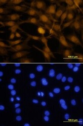
- Immunocytochemistry analysis of OTX2 in immersion fixed NTera‚2 human testicular embryonic carcinoma cell line. Samples were incubated in OTX2 polyclonal antibody (Product # PA5-39887) using a dilution of 10 µg/mL for 3 hours at room temperature followed by NorthernLights™ 557-conjugated Anti-Goat IgG Secondary Antibody (yellow, upper panel) and counterstained with DAPI (blue, lower panel).

- Submitted by
- Invitrogen Antibodies (provider)
- Main image
- Experimental details
- Immunocytochemistry analysis of OTX2 in immersion fixed NTera‚2 human testicular embryonic carcinoma cell line. Samples were incubated in OTX2 polyclonal antibody (Product # PA5-39887) using a dilution of 10 µg/mL for 3 hours at room temperature followed by NorthernLights™ 557-conjugated Anti-Goat IgG Secondary Antibody (yellow, upper panel) and counterstained with DAPI (blue, lower panel).

Supportive validation
- Submitted by
- Invitrogen Antibodies (provider)
- Main image

- Experimental details
- Immunohistochemical analysis of OTX2 in immersion fixed paraffin-embedded sections of mouse embryo (14 d.p.c.). Samples were incubated with OTX2 polyclonal antibody (Product # PA5-39887) using a dilution of 1 µg/mL for 1 hour at room temperature followed by Anti-Goat IgG VisUCyte™ HRP Polymer Antibody. Before incubation with the primary antibody, tissue was subjected to heat-induced epitope retrieval using Antigen Retrieval Reagent-Basic . Tissue was stained using DAB (brown) and counterstained with hematoxylin (blue). Specific staining was localized to developing nervous system.
