Explore
 Validate
 Learn
Learn Western blot
 ELISA
ELISA Immunocytochemistry
Antibody data
- Antibody Data
- Antigen structure
- References [5]
- Comments [0]
- Validations
- Immunocytochemistry [2]
- Immunohistochemistry [1]
- Flow cytometry [2]
- Other assay [2]
Submit
Validation data
Reference
Comment
Report error
- Product number
- MA5-15854 - Provider product page

- Provider
- Invitrogen Antibodies
- Product name
- OTX2 Monoclonal Antibody (1H12C4B5)
- Antibody type
- Monoclonal
- Antigen
- Purifed from natural sources
- Description
- MA5-15854 targets OTX2 in indirect ELISA, FACS, IF, IHC, and WB applications and shows reactivity with Human samples. The MA5-15854 immunogen is purified recombinant fragment of human OTX2 expressed in E. Coli. . MA5-15854 detects OTX2 which has a predicted molecular weight of approximately 32kDa.
- Reactivity
- Human, Mouse, Rat
- Host
- Mouse
- Isotype
- IgG
- Antibody clone number
- 1H12C4B5
- Vial size
- 100 μL
- Concentration
- Conc. not determined
- Storage
- Store at 4°C short term. For long term storage, store at -20°C, avoiding freeze/thaw cycles.
Submitted references Generation of human induced pluripotent stem cell-derived cerebral organoids for cellular and molecular characterization.
Constitutive activation of canonical Wnt signaling disrupts choroid plexus epithelial fate.
Co-activation of Sonic hedgehog and Wnt signaling in murine retinal precursor cells drives ocular lesions with features of intraocular medulloepithelioma.
Patient-derived iPSC-cerebral organoid modeling of the 17q11.2 microdeletion syndrome establishes CRLF3 as a critical regulator of neurogenesis.
Retinal inputs signal astrocytes to recruit interneurons into visual thalamus.
Anastasaki C, Wilson AF, Chen AS, Wegscheid ML, Gutmann DH
STAR protocols 2022 Mar 18;3(1):101173
STAR protocols 2022 Mar 18;3(1):101173
Constitutive activation of canonical Wnt signaling disrupts choroid plexus epithelial fate.
Parichha A, Suresh V, Chatterjee M, Kshirsagar A, Ben-Reuven L, Olender T, Taketo MM, Radosevic V, Bobic-Rasonja M, Trnski S, Holtzman MJ, Jovanov-Milosevic N, Reiner O, Tole S
Nature communications 2022 Feb 2;13(1):633
Nature communications 2022 Feb 2;13(1):633
Co-activation of Sonic hedgehog and Wnt signaling in murine retinal precursor cells drives ocular lesions with features of intraocular medulloepithelioma.
Dottermusch M, Sumisławski P, Krevet J, Middelkamp M, Voß H, Siebels B, Bartsch H, Sotlar K, Meyer P, Frank S, Korshunov A, Glatzel M, Schüller U, Neumann JE
Oncogenesis 2021 Nov 16;10(11):78
Oncogenesis 2021 Nov 16;10(11):78
Patient-derived iPSC-cerebral organoid modeling of the 17q11.2 microdeletion syndrome establishes CRLF3 as a critical regulator of neurogenesis.
Wegscheid ML, Anastasaki C, Hartigan KA, Cobb OM, Papke JB, Traber JN, Morris SM, Gutmann DH
Cell reports 2021 Jul 6;36(1):109315
Cell reports 2021 Jul 6;36(1):109315
Retinal inputs signal astrocytes to recruit interneurons into visual thalamus.
Su J, Charalambakis NE, Sabbagh U, Somaiya RD, Monavarfeshani A, Guido W, Fox MA
Proceedings of the National Academy of Sciences of the United States of America 2020 Feb 4;117(5):2671-2682
Proceedings of the National Academy of Sciences of the United States of America 2020 Feb 4;117(5):2671-2682
No comments: Submit comment
Supportive validation
- Submitted by
- Invitrogen Antibodies (provider)
- Main image

- Experimental details
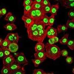
- Immunofluorescence analysis of HepG2 cells using OTX2 monoclonal antibody (Product # MA5-15854) (Green). Red: actin filaments have been labeled with phalloidin.

- Submitted by
- Invitrogen Antibodies (provider)
- Main image
- Experimental details
- Immunofluorescence analysis of HepG2 cells using OTX2 monoclonal antibody (Product # MA5-15854) (Green). Red: actin filaments have been labeled with phalloidin.

Supportive validation
- Submitted by
- Invitrogen Antibodies (provider)
- Main image
- Experimental details
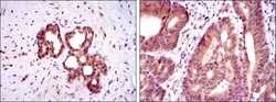
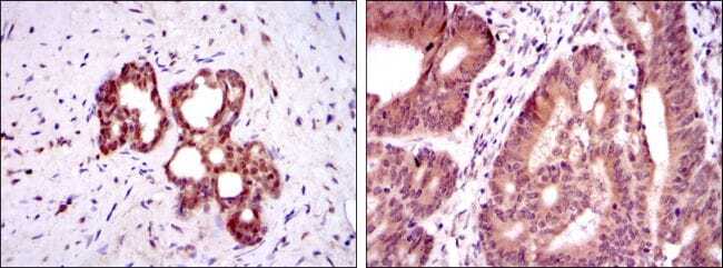
- Immunohistochemical analysis of paraffin-embedded prostate tissues (left) and colon cancer tissues (right) using OTX2 monoclonal antibody (Product # MA5-15854) followed with DAB staining.
Supportive validation
- Submitted by
- Invitrogen Antibodies (provider)
- Main image
- Experimental details
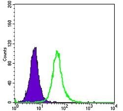
- Flow cytometric analysis of HepG2 cells using OTX2 monoclonal antibody (Product # MA5-15854) (green) and negative control (purple).

- Submitted by
- Invitrogen Antibodies (provider)
- Main image

- Experimental details
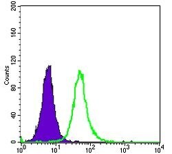
- Flow cytometric analysis of HepG2 cells using OTX2 monoclonal antibody (Product # MA5-15854) (green) and negative control (purple).
Supportive validation
- Submitted by
- Invitrogen Antibodies (provider)
- Main image
- Experimental details
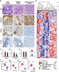
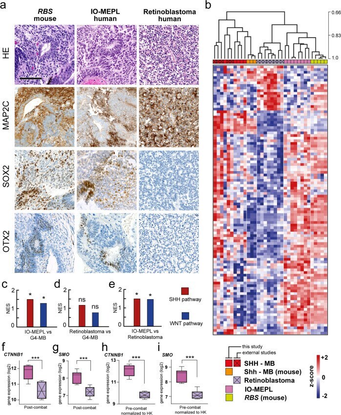
- Fig. 6 Morphological, immunohistochemical, and molecular features of Rax - creER T2 ::Ctnnb1(ex3) fl/+ SmoM2 fl/+ ( RBS ) ocular lesions match with IO-MEPL as opposed to retinoblastoma. a Morphology of murine RBS lesion after tamoxifen administration on day E8.5 (left column) compared to IO-MEPL (middle column) and human retinoblastoma (right column). Flexner-Wintersteiner-Rosettes in retinoblastoma were smaller in comparison to the pseudostratified rosettes of IO-MEPL and RBS (first row). IO-MEPL and RBS stained strongly for MAP2C, while rosette structures consistently lacked staining signal. In contrast, retinoblastoma displayed strong MAP2C positivity with most prominent staining intensity towards the luminal surfaces of rosettes (second row). SOX2-positive cells were found in IO-MEPL and RBS in a scattered distribution with occasional accentuation of staining intensity in association with rosettes or rosette-like structures, while SOX2 staining was negative in Retinoblastoma (third row). OTX2-positive cells were occasionally found in IO-MEPL in association with rosettes or rosette-like structures and in RBS in the residual retina and NBL-like rosettes. In contrast, retinoblastoma stained negative for OTX2 (fourth row). Scale bar is 100 um. b Hierarchical clustering based on cross-species harmonized gene expression data of this study, GSE30074 [] and GSE172170 []. Ocular lesions of RBS mice formed a distinct cluster with closest similarity to human IO-MEPLs, but not human
- Submitted by
- Invitrogen Antibodies (provider)
- Main image
- Experimental details
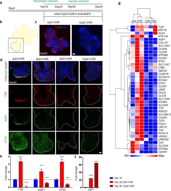
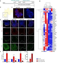
- Fig. 10 High activation of the canonical Wnt pathway in hESC-derived organoids causes suppression of ChPe markers. a A schematic presentation of the protocol for the treatment of organoids with the canonical Wnt agonist CHIR. b , c The ChP marker TTR is present in the periphery of a day 25 organoid exposed to 3 muM, but not when exposed to 12 muM CHIR, from day 18. c N = 4 organoids (3 muM CHIR) and 5 organoids (12 muM CHIR treated); were used for immunohistochemistry analysis. d High magnification images (boxed region shown in b) of serial sections of a day 40 organoid exposed to 3 muM reveal the presence of TTR, AQP1, and OTX2 in the periphery. This labeling is reduced or undetectable upon exposure to higher concentrations of CHIR (6/9/12 muM). d N = 14 organoids (3 muM CHIR), 9 organoids (6 muM CHIR), 5 organoids (9 muM CHIR treated), and 14 organoids (12 muM CHIR treated) from 2 independent experiments were used for analysis (each organoid was considered as an independent biological replicate). Additional examples are provided in Supplementary Fig. S10 . e q-PCR analysis shows that the expression of the ChP markers TTR, AQP1 , and OTX2 is upregulated in organoids at day 30 organoids exposed to 3 muM CHIR from day 18. This increased expression is lost upon treatment with 12 muM CHIR for the same period. ( f ) LEF1 expression displays a stepwise increase upon 3 muM and 12 muM CHIR treatment, consistent with increased activation of the canonical Wnt pathway. Bar graphs ( e ,