 Explore
 Validate
 Learn
Learn Western blot
 ELISA
ELISAAntibody data
- Antibody Data
- Antigen structure
- References [0]
- Comments [0]
- Validations
- Western blot [4]
- Immunocytochemistry [2]
- Immunohistochemistry [2]
Submit
Validation data
Reference
Comment
Report error
- Product number
- MA5-15855 - Provider product page

- Provider
- Invitrogen Antibodies
- Product name
- OTX2 Monoclonal Antibody (1H12G8B2)
- Antibody type
- Monoclonal
- Antigen
- Purifed from natural sources
- Description
- MA5-15855 targets OTX2 in indirect ELISA, IF, IHC, and WB applications and shows reactivity with Human samples. The MA5-15855 immunogen is purified recombinant fragment of human OTX2 expressed in E. Coli. . MA5-15855 detects OTX2 which has a predicted molecular weight of approximately 32kDa.
- Reactivity
- Human, Mouse, Rat
- Host
- Mouse
- Isotype
- IgG
- Antibody clone number
- 1H12G8B2
- Vial size
- 100 µL
- Concentration
- Conc. Not Determined
- Storage
- Store at 4°C short term. For long term storage, store at -20°C, avoiding freeze/thaw cycles.
No comments: Submit comment
Supportive validation
- Submitted by
- Invitrogen Antibodies (provider)
- Main image

- Experimental details
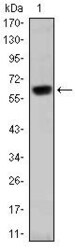
- Western blot analysis of OTX2 using a OTX2 monoclonal antibody (Product # MA5-15855) against a human OTX2 (AA: 40-297) recombinant protein.

- Submitted by
- Invitrogen Antibodies (provider)
- Main image
- Experimental details
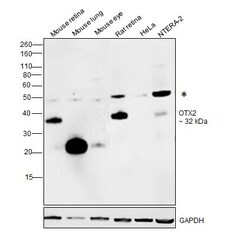
- Western blot was performed using Anti-OTX2 Monoclonal Antibody (1H12G8B2) (Product # MA5-15855) and a 32 kDa band corresponding to OTX2 was observed in mouse retina and rat retina as compared to other tissues. A band at ~25kDa corresponding to circulating tissue IgG was observed in the Mouse tissue lysates. Tissue extracts (30 µg lysate) of Mouse retina (Lane 1), Mouse lung (Lane 2), Mouse eye (Lane 3), Rat retina (Lane 4), modified whole cell extract (1% SDS) of HeLa (Lane 5) and NTERA-2 (Lane 6) were electrophoresed using Novex® NuPAGE® 12 % Bis-Tris gel (Product # NP0342BOX). Resolved proteins were then transferred onto a nitrocellulose membrane (Product # IB23001) by iBlot® 2 Dry Blotting System (Product # IB21001). The blot was probed with the primary antibody (1:1000 dilution) and detected by chemiluminescence with Goat anti-Mouse IgG (H+L) Superclonal™ Recombinant Secondary Antibody, HRP (Product # A28177, 1:4000 dilution) using the iBright FL 1000 (Product # A32752). Chemiluminescent detection was performed using Novex® ECL Chemiluminescent Substrate Reagent Kit (Product # WP20005).

- Submitted by
- Invitrogen Antibodies (provider)
- Main image
- Experimental details
- Western blot analysis of OTX2 using a OTX2 monoclonal antibody (Product # MA5-15855) against a human OTX2 (AA: 40-297) recombinant protein.

- Submitted by
- Invitrogen Antibodies (provider)
- Main image

- Experimental details
- Western blot analysis of OTX2 using OTX2 monoclonal antibody (Product # MA5-15855) in HepG2 (1), Jurkat (2), and NTERA-2 (3) cell lysate.

Supportive validation
- Submitted by
- Invitrogen Antibodies (provider)
- Main image

- Experimental details
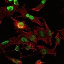
- Immunofluorescence analysis of U251 cells using OTX2 monoclonal antibody (Product # MA5-15855) (Green). Red: actin filaments have been labeled with phalloidin.

- Submitted by
- Invitrogen Antibodies (provider)
- Main image
- Experimental details
- Immunofluorescence analysis of U251 cells using OTX2 monoclonal antibody (Product # MA5-15855) (Green). Red: actin filaments have been labeled with phalloidin.

Supportive validation
- Submitted by
- Invitrogen Antibodies (provider)
- Main image

- Experimental details
- Immunohistochemical analysis of paraffin-embedded colon tissues (left) and colon cancer tissues (right) using OTX2 monoclonal antibody (Product # MA5-15855) followed with DAB staining.

- Submitted by
- Invitrogen Antibodies (provider)
- Main image

- Experimental details
- Immunohistochemical analysis of paraffin-embedded stomach tissues (left) and brain tissues (right) using OTX2 monoclonal antibody (Product # MA5-15855) followed with DAB staining.
