Explore
 Validate
 Learn
Learn Western blot
Antibody data
- Antibody Data
- Antigen structure
- References [1]
- Comments [0]
- Validations
- Western blot [3]
- Immunocytochemistry [2]
- Immunohistochemistry [5]
Submit
Validation data
Reference
Comment
Report error
- Product number
- TA500072 - Provider product page

- Provider
- OriGene
- Proper citation
- OriGene Cat#TA500072, RRID:AB_2257799
- Product name
- alpha-actinin (Actinin alpha 1) mouse monoclonal antibody, clone OTI7A4 (formerly 7A4)
- Antibody type
- Monoclonal
- Description
- alpha-actinin (Actinin alpha 1) mouse monoclonal antibody, clone OTI7A4 (formerly 7A4)
- Host
- Mouse
- Conjugate
- Unconjugated
- Epitope
- ACTN1
- Isotype
- IgG
- Antibody clone number
- OTI7A4
- Vial size
- 100 µl
- Concentration
- 0.88 mg/ml
Submitted references Involvement of ubiquitin-conjugating enzyme E2C in proliferation and invasion of prostate carcinoma cells.
Shuliang S, Lei C, Guangwu J, Changjie L
Oncology research 2013;21(3):121-7
Oncology research 2013;21(3):121-7
No comments: Submit comment
Supportive validation
- Submitted by
- OriGene (provider)
- Main image
- Experimental details
- HEK293T cells were transfected with the pCMV6-ENTRY control (Left lane) or pCMV6-ENTRY ACTN1 (RC204924, Right lane) cDNA for 48 hrs and lysed. Equivalent amounts of cell lysates (5 ug per lane) were separated by SDS-PAGE and immunoblotted with anti-ACTN1.
- Validation comment
- WB
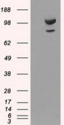
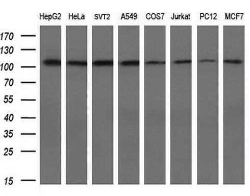
- Submitted by
- OriGene (provider)
- Main image

- Experimental details
- Western blot analysis of extracts (10ug) from 8 different cell lines by using anti-ACTN1 monoclonal antibody at 1:200 dilution.
- Validation comment
- WB
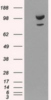
- Submitted by
- OriGene (provider)
- Main image
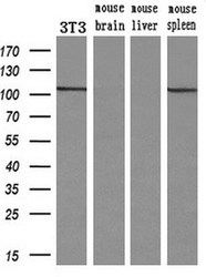
- Experimental details
- Western blot analysis of extracts (10ug) from a mouse cell line and 3 different mouse tissues by using anti-ACTN1 monoclonal antibody.(1:200)
- Validation comment
- WB

Supportive validation
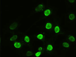
- Submitted by
- OriGene (provider)
- Main image

- Experimental details
- Anti-ACTN1 mouse monoclonal antibody (TA500072) immunofluorescent staining of Hela cells transiently transfected by pCMV6-ENTRY ACTN1(RC204924).
- Validation comment
- IF
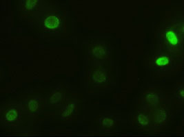
- Submitted by
- OriGene (provider)
- Main image

- Experimental details
- Immunofluorescent staining of HeLa cells using anti-ACTN1 mouse monoclonal antibody (TA500072).
- Validation comment
- IF
Supportive validation
- Submitted by
- OriGene (provider)
- Main image
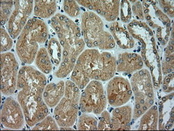
- Experimental details
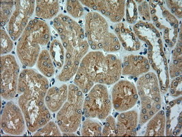
- Immunohistochemical staining of paraffin-embedded Human Kidney tissue within the normal limits using anti-ACTN1 mouse monoclonal antibody. (Heat-induced epitope retrieval by 10mM citric buffer, pH6.0, 100C for 10min, TA500072)
- Validation comment
- IHC
- Submitted by
- OriGene (provider)
- Main image
- Experimental details
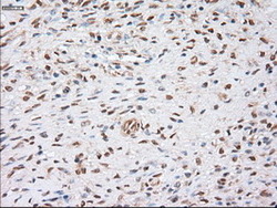
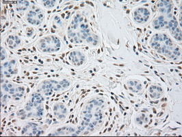
- Immunohistochemical staining of paraffin-embedded Human Ovary tissue within the normal limits using anti-ACTN1 mouse monoclonal antibody. (Heat-induced epitope retrieval by 10mM citric buffer, pH6.0, 100C for 10min, TA500072)
- Validation comment
- IHC
- Submitted by
- OriGene (provider)
- Main image

- Experimental details
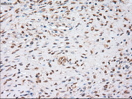
- Immunohistochemical staining of paraffin-embedded Human liver tissue within the normal limits using anti-ACTN1 mouse monoclonal antibody. (Heat-induced epitope retrieval by 10mM citric buffer, pH6.0, 100C for 10min, TA500072)
- Validation comment
- IHC

- Submitted by
- OriGene (provider)
- Main image
- Experimental details
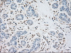
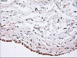
- Immunohistochemical staining of paraffin-embedded Human breast tissue within the normal limits using anti-ACTN1 mouse monoclonal antibody. (Heat-induced epitope retrieval by 10mM citric buffer, pH6.0, 100C for 10min, TA500072)
- Validation comment
- IHC
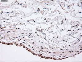
- Submitted by
- OriGene (provider)
- Main image
- Experimental details
- Immunohistochemical staining of paraffin-embedded Human bladder tissue within the normal limits using anti-ACTN1 mouse monoclonal antibody. (Heat-induced epitope retrieval by 10mM citric buffer, pH6.0, 100C for 10min, TA500072)
- Validation comment
- IHC