Antibody data
- Antibody Data
- Antigen structure
- References [2]
- Comments [0]
- Validations
- Western blot [6]
- Immunohistochemistry [1]
- Other assay [1]
Submit
Validation data
Reference
Comment
Report error
- Product number
- PA5-27164 - Provider product page
- Provider
- Invitrogen Antibodies
- Product name
- HTR2C Polyclonal Antibody
- Antibody type
- Polyclonal
- Antigen
- Synthetic peptide
- Description
- Recommended positive controls: IMR32, SK-N-AS, mouse placenta, rat brain. Predicted reactivity: Mouse (100%), Rat (100%), Dog (100%), Chimpanzee (100%). Store product as a concentrated solution. Centrifuge briefly prior to opening the vial.
- Reactivity
- Human, Mouse, Rat
- Host
- Rabbit
- Isotype
- IgG
- Vial size
- 100 µL
- Concentration
- 0.68 mg/mL
- Storage
- Store at 4°C short term. For long term storage, store at -20°C, avoiding freeze/thaw cycles.
Submitted references Effects of 5-Hydroxytryptamine Class 2 Receptor Antagonists on Bronchoconstriction and Pulmonary Remodeling Processes.
5-HT2B receptor antagonists attenuate myofibroblast differentiation and subsequent fibrotic responses in vitro and in vivo.
Löfdahl A, Wenglén C, Rydell-Törmänen K, Westergren-Thorsson G, Larsson-Callerfelt AK
The American journal of pathology 2018 May;188(5):1113-1119
5-HT2B receptor antagonists attenuate myofibroblast differentiation and subsequent fibrotic responses in vitro and in vivo.
Löfdahl A, Rydell-Törmänen K, Müller C, Martina Holst C, Thiman L, Ekström G, Wenglén C, Larsson-Callerfelt AK, Westergren-Thorsson G
Physiological reports 2016 Aug;4(15)
No comments: Submit comment
Supportive validation
- Submitted by
- Invitrogen Antibodies (provider)
- Main image
- Experimental details
- Western blot analysis of 5HT2C Receptor using 30 µg of A431 lysate. Samples were loaded onto a 10% SDS-PAGE gel and probed with a 5HT2C Receptor polyclonal antibody (Product # PA5-27164) at a dilution of 1:1000.
- Submitted by
- Invitrogen Antibodies (provider)
- Main image
- Experimental details
- Western Blot analysis of HTR2C was performed by separating 50 µg of mouse tissue extract by 10% SDS-PAGE. Proteins were transferred to a membrane and probed with a HTR2C Polyclonal Antibody (Product # PA5-27164) at a dilution of 1:500.
- Submitted by
- Invitrogen Antibodies (provider)
- Main image
- Experimental details
- Western Blot using HTR2C Polyclonal Antibody (Product # PA5-27164). Various whole cell extracts (30 µg) were separated by 10% SDS-PAGE, and the membrane was blotted with HTR2C Polyclonal Antibody (Product # PA5-27164) diluted at 1:500. The HRP-conjugated anti-rabbit IgG antibody was used to detect the primary antibody.
- Submitted by
- Invitrogen Antibodies (provider)
- Main image
- Experimental details
- Western Blot analysis of HTR2C was performed by separating 50 µg of rat tissue extract by 10% SDS-PAGE. Proteins were transferred to a membrane and probed with a HTR2C Polyclonal Antibody (Product # PA5-27164) at a dilution of 1:1,000.
- Submitted by
- Invitrogen Antibodies (provider)
- Main image
- Experimental details
- Western Blot analysis of HTR2C was performed by separating 30 µg of various whole cell extracts by 10% SDS-PAGE. Proteins were transferred to a membrane and probed with a HTR2C Polyclonal Antibody (Product # PA5-27164) at a dilution of 1:500.
- Submitted by
- Invitrogen Antibodies (provider)
- Main image
- Experimental details
- Western blot was performed using Anti-HTR2C Polyclonal Antibody (Product # PA5-27164) and a 60 kDa band corresponding to HTR2C was observed across the panel tested except for Mouse and Rat Eye which are reported to be negative. Membrane enriched extracts (30 µg lysate) of SH-SY5Y (Lane 1), U-87 MG (Lane 2), Neuro-2a (Lane 3), IMR-32 (Lane 4), A-431 (Lane 5), Mouse Brain (Lane 6), Mouse Eye (Lane 7), Rat Brain (Lane 8), Rat Eye (Lane 9) were electrophoresed using NuPAGE™ 4-12% Bis-Tris Protein Gel (Product # NP0321BOX). Resolved proteins were then transferred onto a Nitrocellulose membrane (Product # IB23001) by iBlot® 2 Dry Blotting System (Product # IB21001). The blot was probed with the primary antibody (1:500 dilution) and detected by chemiluminescence with Goat anti-Rabbit IgG (H+L) Superclonal™ Recombinant Secondary Antibody, HRP (Product # A27036,1:4000 dilution) using the iBright FL 1000 (Product # A32752). Chemiluminescent detection was performed using Novex® ECL Chemiluminescent Substrate Reagent Kit (Product # WP20005).
Supportive validation
- Submitted by
- Invitrogen Antibodies (provider)
- Main image
- Experimental details
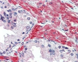
- Immunohistochemical analysis of paraffin-embedded human prostate, using 5HT2C Receptor (Product # PA5-27164) antibody (10 µg/mL). Antigen Retrieval: Citrate buffer, pH 6.0, 10 min.
Supportive validation
- Submitted by
- Invitrogen Antibodies (provider)
- Main image
- Experimental details
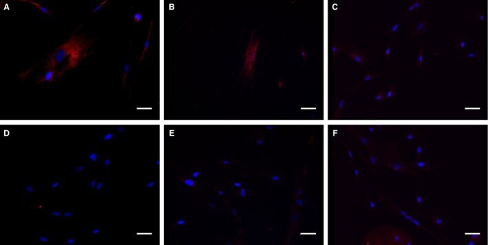
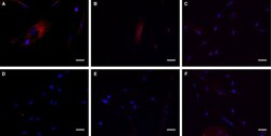
- Figure 1 HFL -1 expression of 5- HT 2 receptors. Identification of 5- HT 2A (A), 5- HT 2B (B), and 5- HT 2C (C) receptors on cultured HFL -1 cells. Isotype-matched negative controls for 5- HT 2A (D), 5- HT 2B (E), and 5- HT 2C (F) receptor antibodies showed no positive receptor staining. Anti-5- HT 2 receptor antibodies (red), cell nuclear staining with DAPI (blue). Scale bars represent 40 mu m.