Explore
 Validate
 Learn
Learn Western blot
 Immunohistochemistry
Antibody data
- Antibody Data
- Antigen structure
- References [1]
- Comments [0]
- Validations
- Immunohistochemistry [1]
- Flow cytometry [2]
- Other assay [2]
Submit
Validation data
Reference
Comment
Report error
- Product number
- PA5-25750 - Provider product page

- Provider
- Invitrogen Antibodies
- Product name
- RASGRP1 Polyclonal Antibody
- Antibody type
- Polyclonal
- Antigen
- Synthetic peptide
- Description
- This antibody is predicted to react with mouse and rat based on sequence homology.
- Reactivity
- Human
- Host
- Rabbit
- Isotype
- IgG
- Vial size
- 400 μL
- Concentration
- 2 mg/mL
- Storage
- Store at 4°C short term. For long term storage, store at -20°C, avoiding freeze/thaw cycles.
Submitted references Loss of RASGRP1 in humans impairs T-cell expansion leading to Epstein-Barr virus susceptibility.
Winter S, Martin E, Boutboul D, Lenoir C, Boudjemaa S, Petit A, Picard C, Fischer A, Leverger G, Latour S
EMBO molecular medicine 2018 Feb;10(2):188-199
EMBO molecular medicine 2018 Feb;10(2):188-199
No comments: Submit comment
Supportive validation
- Submitted by
- Invitrogen Antibodies (provider)
- Main image
- Experimental details
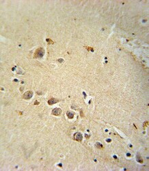
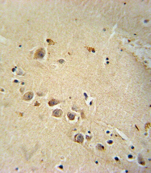
- Immunohistochemistry analysis of RASGRP1 in formalin fixed and paraffin embedded human brain tissue. Samples were incubated with RASGRP1 polyclonal antibody (Product # PA5-25750) followed by peroxidase conjugation of the secondary antibody and DAB staining. This data demonstrates the use of this antibody for immunohistochemistry. Clinical relevance has not been evaluated.
Supportive validation
- Submitted by
- Invitrogen Antibodies (provider)
- Main image

- Experimental details
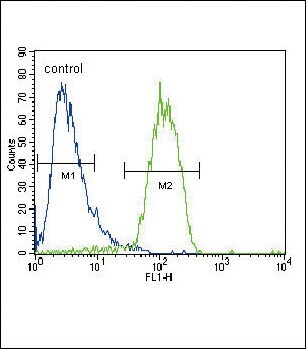
- Flow cytometry analysis of HepG2 cells using a RASGRP1 polyclonal antibody (Product # PA5-25750) (right) compared to a negative control cell (left) at a dilution of 1:10-50, followed by a FITC-conjugated goat anti-rabbit antibody
- Submitted by
- Invitrogen Antibodies (provider)
- Main image
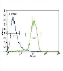
- Experimental details
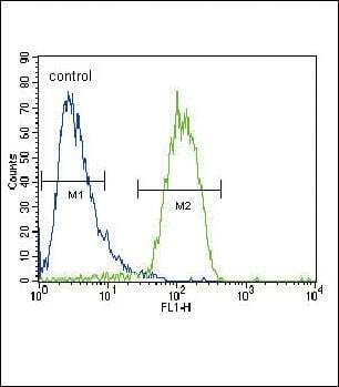
- Flow cytometry of RASGRP1 in HepG2 cells (right histogram). Samples were incubated with RASGRP1 polyclonal antibody (Product # PA5-25750) followed by FITC-conjugated goat-anti-rabbit secondary antibody. Negative control cell (left histogram).
Supportive validation
- Submitted by
- Invitrogen Antibodies (provider)
- Main image
- Experimental details
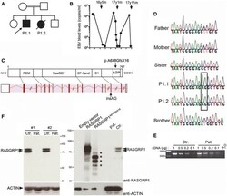
- Figure 1 Identification of a homozygous loss-of-function mutation in RASGRP1 in two siblings with Hodgkin lymphoma and defective immunity to EBV Pedigree of the family in which the c.1910_1911insAG mutation in RASGRP1 was identified. The arrow indicates the proband (P1.1) who was analyzed by WES. EBV load in the blood of patient P1.1 is shown as the number of EBV copies detected by PCR at different time points (black circles). Arrows correspond to the anti-CD20/rituximab treatments received by the patient with the age (year, y; month, m) of patient at the time of the treatment. Schematic representation of intron-exon organization of the RASGRP1 gene and its correspondence at protein level with the different domains of RASGRP1 shown: the Ras exchanger motif (REM), the Ras-guanine exchange factor (RasGEF), the EF-hand, the C1, and the bZIP domains. The mutation is indicated by an arrow at gene and protein levels. DNA electropherograms of the family showing the g.38786931_38786932insAG mutation in P1.1 and P1.2 that is shown in the box. Expression of RASGRP1 transcript in T-cell blasts of healthy control and the patient P1.1 (Pat.). The relative expression of full-length RASGRP1 transcript was examined by qRT-PCR in T-cell blasts of a healthy control and P1.1. Fourfold serial dilutions of cDNAs (1, 0.5, 0.25, and 0.12) were used for amplification of each transcript after quantitation. Base pair markers are shown on the left. PCR products were verified by sequencing showing th

- Submitted by
- Invitrogen Antibodies (provider)
- Main image

- Experimental details
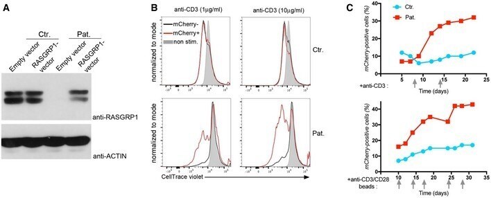
- Figure 5 RASGRP1 is required for T-cell expansion in response to CD3-TCR activation Rescue of T-cell proliferation by expression of wild-type RASGRP1 in RASGRP1-deficient T cells. Immunoblots for RASGRP1 and actin as a loading control of control (Ctr.) and RASGRP1-deficient T cells of patient P1.1 (Pat.) transduced by empty or wild-type RASGRP1-containing vector with mCherry gene reporter at day 14 of culture. Proliferation of control (Ctr.) and RASGRP1-deficient T cells of patient P1.1 (Pat.) transduced by empty or wild-type RASGRP1-containing vector. Representative histograms of CellTrace violet dye dilution showing the cell divisions of mCherry-positive (mCherry + ) and mCherry-negative (mCherry - ) cells after stimulation with two concentrations of anti-CD3 antibody, with histograms in gray corresponding to unstimulated cells (unstim.). Curves of the percentage of mCherry-positive transduced cells after repeated stimulations indicated by gray arrows with anti-CD3 (upper panels) or anti-CD3/CD28 (lower panels) antibodies at different days in long-term expansions. Data information: Representative data from two independent experiments. Source data are available online for this figure.