Explore
 Validate
 Learn
Learn Western blot
 Other assay
Antibody data
- Antibody Data
- Antigen structure
- References [5]
- Comments [0]
- Validations
- Other assay [9]
Submit
Validation data
Reference
Comment
Report error
- Product number
- PA5-75342 - Provider product page

- Provider
- Invitrogen Antibodies
- Product name
- MC1R Polyclonal Antibody
- Antibody type
- Polyclonal
- Antigen
- Synthetic peptide
- Description
- The antibody was affinity-purified from rabbit antiserum by affinity-chromatography using epitope-specific immunogen and the purity is > 95% (by SDS-PAGE).
- Reactivity
- Human, Mouse, Rat
- Host
- Rabbit
- Isotype
- IgG
- Vial size
- 100 μL
- Concentration
- 1 mg/mL
- Storage
- Store at 4°C short term. For long term storage, store at -20°C, avoiding freeze/thaw cycles.
Submitted references BMS-470539 Attenuates Oxidative Stress and Neuronal Apoptosis via MC1R/cAMP/PKA/Nurr1 Signaling Pathway in a Neonatal Hypoxic-Ischemic Rat Model.
Visualized podocyte-targeting and focused ultrasound responsive glucocorticoid nano-delivery system against immune-associated nephropathy without glucocorticoid side effect.
Melanocortin 1 receptor attenuates early brain injury following subarachnoid hemorrhage by controlling mitochondrial metabolism via AMPK/SIRT1/PGC-1α pathway in rats.
Activation of MC1R with BMS-470539 attenuates neuroinflammation via cAMP/PKA/Nurr1 pathway after neonatal hypoxic-ischemic brain injury in rats.
Activation of Melanocortin 1 Receptor Attenuates Early Brain Injury in a Rat Model of Subarachnoid Hemorrhage viathe Suppression of Neuroinflammation through AMPK/TBK1/NF-κB Pathway in Rats.
Yu S, Doycheva DM, Gamdzyk M, Gao Y, Guo Y, Travis ZD, Tang J, Chen WX, Zhang JH
Oxidative medicine and cellular longevity 2022;2022:4054938
Oxidative medicine and cellular longevity 2022;2022:4054938
Visualized podocyte-targeting and focused ultrasound responsive glucocorticoid nano-delivery system against immune-associated nephropathy without glucocorticoid side effect.
Fan K, Zeng L, Guo J, Xie S, Yu Y, Chen J, Cao J, Xiang Q, Zhang S, Luo Y, Deng Q, Zhou Q, Zhao Y, Hao L, Wang Z, Zhong L
Theranostics 2021;11(6):2670-2690
Theranostics 2021;11(6):2670-2690
Melanocortin 1 receptor attenuates early brain injury following subarachnoid hemorrhage by controlling mitochondrial metabolism via AMPK/SIRT1/PGC-1α pathway in rats.
Xu W, Yan J, Ocak U, Lenahan C, Shao A, Tang J, Zhang J, Zhang JH
Theranostics 2021;11(2):522-539
Theranostics 2021;11(2):522-539
Activation of MC1R with BMS-470539 attenuates neuroinflammation via cAMP/PKA/Nurr1 pathway after neonatal hypoxic-ischemic brain injury in rats.
Yu S, Doycheva DM, Gamdzyk M, Yang Y, Lenahan C, Li G, Li D, Lian L, Tang J, Lu J, Zhang JH
Journal of neuroinflammation 2021 Jan 19;18(1):26
Journal of neuroinflammation 2021 Jan 19;18(1):26
Activation of Melanocortin 1 Receptor Attenuates Early Brain Injury in a Rat Model of Subarachnoid Hemorrhage viathe Suppression of Neuroinflammation through AMPK/TBK1/NF-κB Pathway in Rats.
Xu W, Mo J, Ocak U, Travis ZD, Enkhjargal B, Zhang T, Wu P, Peng J, Li T, Zuo Y, Shao A, Tang J, Zhang J, Zhang JH
Neurotherapeutics : the journal of the American Society for Experimental NeuroTherapeutics 2020 Jan;17(1):294-308
Neurotherapeutics : the journal of the American Society for Experimental NeuroTherapeutics 2020 Jan;17(1):294-308
No comments: Submit comment
Supportive validation
- Submitted by
- Invitrogen Antibodies (provider)
- Main image
- Experimental details
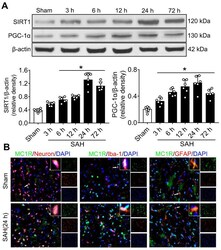
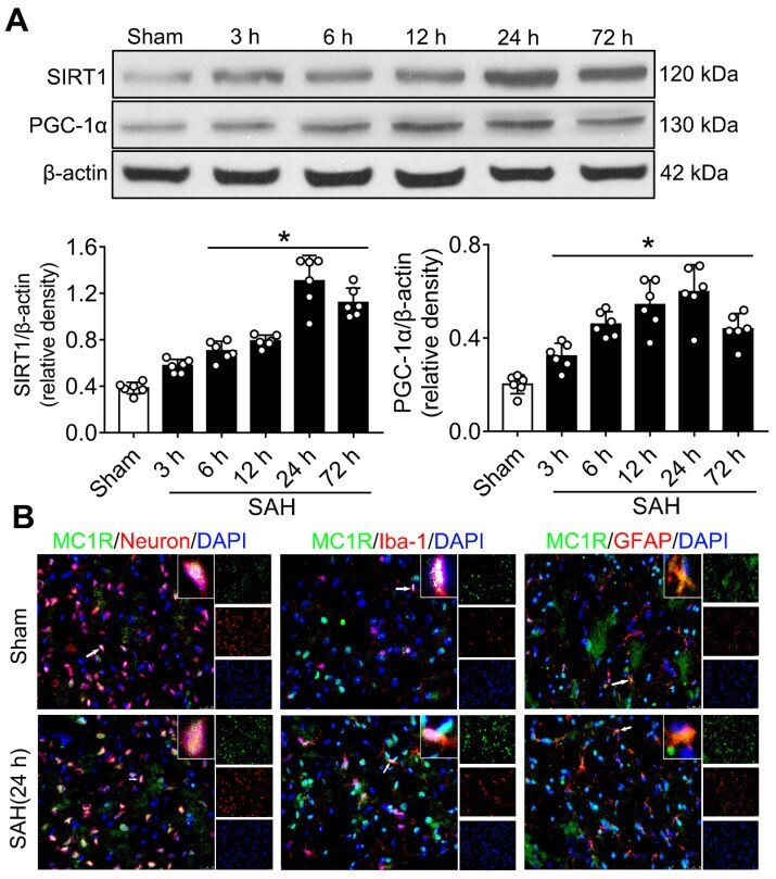
- Figure 1 The expression of MC1R, SIRT1 and PGC-1alpha. (A) Representative western blot images and quantitative analyses of SIRT1 and PGC-1alpha of the time course study in samples obtained from the left hemisphere after SAH; n = 6 for each group. Data were shown as means +- SD, and were compared by 1-way ANOVA followed by the Tukey post-hoc test. * p < 0.05 vs. sham; (B) Representative microphotographs of immune-fluorescence staining, showing colocalization of MC1R (green) with NeuN, iba-1, and GFAP (all red) 24 h in the sham and SAH (24 h) groups. (n = 2 for each group). Scale bar = 50 mum.
- Submitted by
- Invitrogen Antibodies (provider)
- Main image
- Experimental details
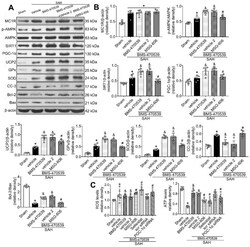
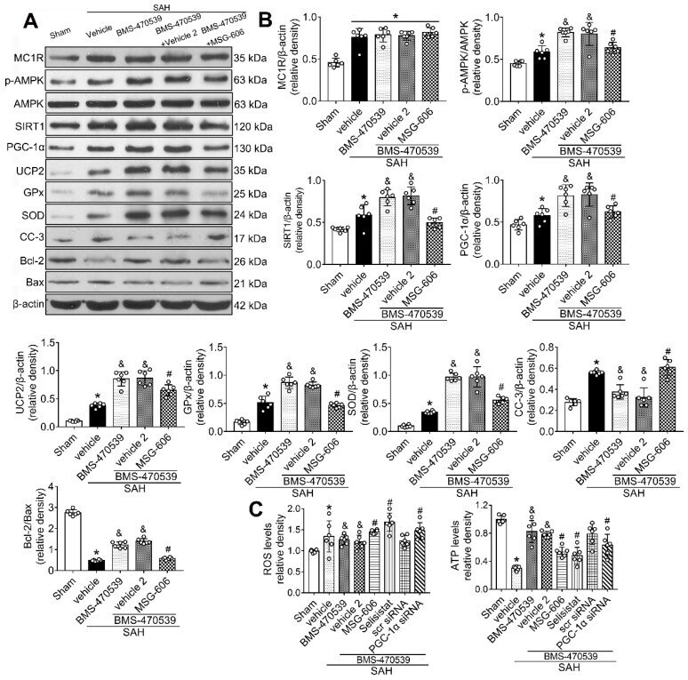
- Figure 5 Inhibition of MC1R with MSG-606 abolished the neuroprotective effects of BMS-470539 at 24 h after SAH. (A) Representative western blot images; (B) Quantitative analyses of MC1R, p-AMPK, SIRT1, PGC-1alpha, UCP2, GPx, SOD, CC-3, Bcl-2, Bax; (C) ROS and ATP levels at 24 h after SAH. n = 6 for each group. Data were shown as means +- SD and compared by 1-way ANOVA followed by the Tukey post-hoc test. * p < 0.05 vs. sham, & p < 0.05 vs. SAH + vehicle, # p < 0.05 vs. SAH + BMS-470539.
- Submitted by
- Invitrogen Antibodies (provider)
- Main image
- Experimental details
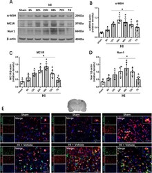
- Fig. 1 Expression levels of alpha-MSH, MC1R, and Nurr1 post-HI. a Representative pictures of Western blot data. b Western blot data showed that the expression levels of alpha-MSH significantly increased at 12 h post-HI, and peaked at 24 h. c , d The endogenous expression levels of MC1R and Nurr1 increased over time and peaked at 48 h post-HI. Data were represented as mean +- SD. Statistical differences between groups were analyzed using one-way ANOVA, followed by Tukey''s post hoc test (* p < 0.05 versus sham, # p < 0.05 versus 6 h HI, @ p < 0.05 versus 24 h HI, & p < 0.05 versus 48 h HI; n = 4 per group). e Representative microphotographs of double immunofluorescence of alpha-MSH (green) with microglia (Iba-1, red), astrocyte (GFAP, red), and neuron (NeuN, red) in the peri-infarcted area at 24 h post-HI. DAPI was stained blue. Merged images showed that alpha-MSH was colocalized with microglia, astrocytes, and neurons. n = 2 per group. Scale bar = 100 mum
- Submitted by
- Invitrogen Antibodies (provider)
- Main image
- Experimental details
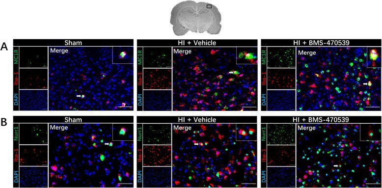
- Fig. 3 Immunofluorescence staining of MC1R and Nurr1 with microglia in the brain at 48 h post-HI. Immunofluorescence staining showed an increase expression of MC1R ( a ) and Nurr1 ( b ) on microglia in the vehicle group when compared with the sham group, and further increased expression of MC1R ( a ) and Nurr1 ( b ) in the BMS-470359 treatment group. Merge showed the colocalization of MC1R and Nurr1 on microglia. Microglia were stained red. MC1R and Nurr1 were stained green. DAPI was stained blue. n = 2 per group. Scale bar = 100 mum
- Submitted by
- Invitrogen Antibodies (provider)
- Main image
- Experimental details
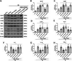
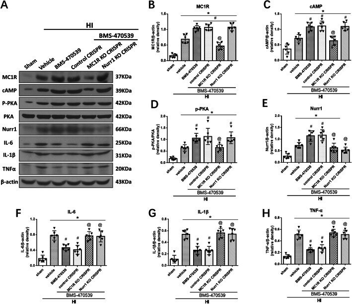
- Fig. 8 MC1R activation on neuroinflammation via cAMP/PKA/Nurr1 signaling pathway at 48 h post-HI. a Representative picture of Western blot data showing bands of the expression levels of MC1R, cAMP, p-PKA, Nurr1, IL-1beta, TNFalpha, and IL-6 either with BMS-470539 treatment alone, BMS-470539 + MC1R KO CRISPR, BMS-470539 + Nurr1 KO CRISPR, and BMS-470539 + control CRISPR groups. b - h Western blot data quantification of bands showed that BMS-470539 treatment significantly increased the expression of MC1R, cAMP, p-PKA, and Nurr1 compared to the HI + vehicle group. Knockout of MC1R using CRISPR significantly decreased MC1R, cAMP, p-PKA, and Nurr1 expression levels compared to the HI + BMS-470539 group or HI + BMS-470539 + control CRISPR group. Furthermore, knockout of Nurr1 using CRISPR significantly decreased the levels of Nurr1, but did not affect MC1R, cAMP, and p-PKA expression compared to the corresponding controls. Activation of MC1R with BMS-470539 showed significantly decreased levels of IL-1beta, TNFalpha, and IL-6, while both treatment groups with CRISPR interventions significantly reversed these effects. Data was represented as mean +- SD. The one-way ANOVA was followed by Tukey''s post hoc test (* p < 0.05 versus sham; # p < 0.05 versus HI + vehicle; @ p < 0.05 HI + BMS-470539 or HI + BMS-470539 + control CRISPR; n = 6 per group)
- Submitted by
- Invitrogen Antibodies (provider)
- Main image
- Experimental details
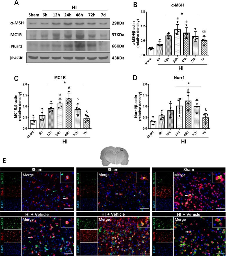
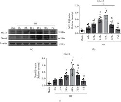
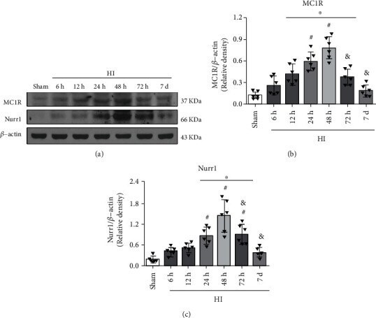
- Figure 1 Temporal expression of endogenous MC1R and Nurr1 in the ipsilateral brain hemisphere post-HI. (a) Representative Western blot bands of the time course. (b) Western blot data showed that the endogenous expression levels of MC1R significantly increased from 12 h reaching peak at 48 h post-HI. (c) Nurr1 expression levels significantly increased over time, reached highest at the 48 h post-HI. n = 6 per group. Data were represented as mean +- SD. * p < 0.05 versus sham, # p < 0.05 versus 6 h HI, and & p < 0.05 versus 48 h HI; one-way ANOVA, Tukey's post hoc test.
- Submitted by
- Invitrogen Antibodies (provider)
- Main image
- Experimental details
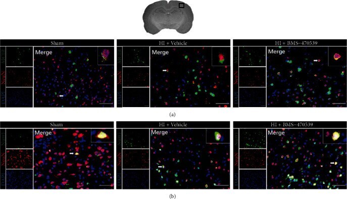
- Figure 3 Immunofluorescence staining showed MC1R and Nurr1 colocalization with neurons in the ipsilateral brain hemisphere at 48 h post-HI. Immunofluorescence staining showed that MC1R (a) and Nurr1 (b) expressions on neurons were seen to be higher in the vehicle-treated pup rats compared to the sham group, and a higher expression of MC1R (a) and Nurr1 (b) on neurons after BMS-470359 treatment compared with vehicle. Merge showed the colocalization of MC1R and Nurr1 on neurons. n = 2 per group. Neurons were stained red. MC1R and Nurr1 were stained green. DAPI was stained blue. Scale bar = 100 mu m.
- Submitted by
- Invitrogen Antibodies (provider)
- Main image
- Experimental details
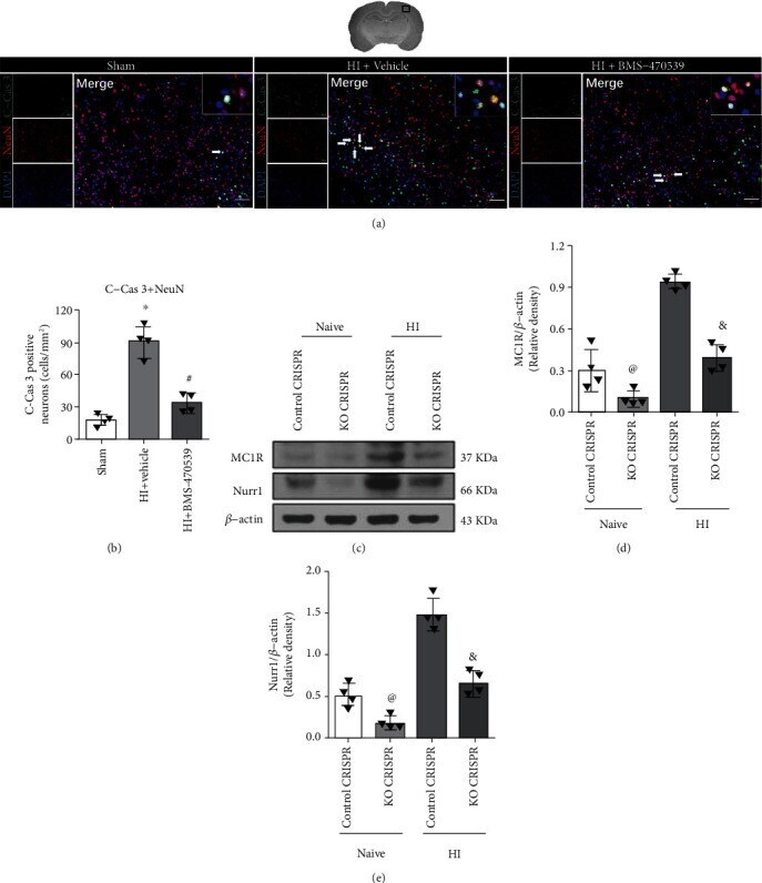
- Figure 4 Effects of BMS-470539 treatment on neuronal apoptosis at 48 h post-HI, and the knockout efficiency of MC1R or Nurr1 knockout CRISPR in naive and HI rats. (a, b) Representative microphotographs and quantitative analysis of C-Cas 3-positive neurons in the ipsilateral hemisphere at 48 h post-HI. The number of C-Cas 3-positive neurons significantly increased in the vehicle group compared with the sham group and BMS-470539 treatment group. n = 4 per group. C-Cas 3 was green. Neurons were red. Blue was for DAPI. Scale bar = 100 mu m. (c, d) Representative Western blot bands and quantitative analysis of MC1R and Nurr1 protein levels in the ipsilateral hemisphere at 48 h post-HI. The expression of MC1R or Nurr1 was significantly reduced by MC1R or Nurr1 knockout CRISPR in naive and HI rats. n = 4 per group (data were represented as mean +- SD; * p < 0.05 versus sham; # p
- Submitted by
- Invitrogen Antibodies (provider)
- Main image
- Experimental details
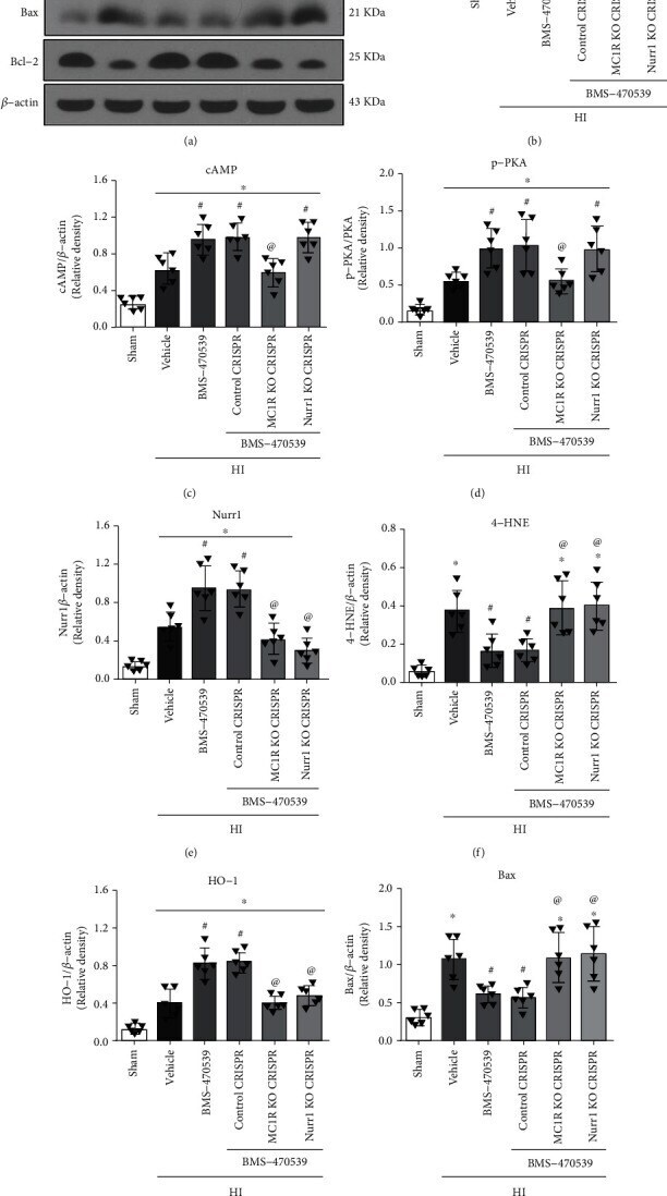
- Figure 5 BMS-470539 treatment exerted its antioxidative stress and antiapoptosis effects via MC1R/cAMP/PKA/Nurr1 signaling pathway at 48 h post-HI. (a) Representative Western blot bands. (b-i) Quantification of MC1R, cAMP, p-PKA, Nurr1, 4-HNE, HO-1, Bax, and Bcl-2 expression levels in the ipsilateral hemisphere at 48 h post-HI. BMS-470539 treatment significantly increased the protein levels of MC1R, cAMP, p-PKA, Nurr1, HO-1, and Bcl-2 but significantly decreased the expression of 4-HNE and Bax compared to the HI+vehicle group. Knockout of MC1R or Nurr1 with CRISPR interventions significantly abolished such effects of BMS-470539. n = 6 per group. Data was represented as mean +- SD. * p < 0.05 versus sham, # p < 0.05 versus HI+vehicle, @ p < 0.05 HI+BMS-470539 or HI+BMS-470539+control CRISPR; one-way ANOVA, Tukey's post hoc test.