Explore
 Validate
 Learn
Learn Western blot
Antibody data
- Antibody Data
- Antigen structure
- References [0]
- Comments [0]
- Validations
- Western blot [3]
- Immunocytochemistry [1]
- Immunohistochemistry [1]
Submit
Validation data
Reference
Comment
Report error
- Product number
- AMR-020-200UL - Provider product page

- Provider
- Invitrogen Antibodies
- Product name
- MC1 Receptor Polyclonal Antibody
- Antibody type
- Polyclonal
- Antigen
- Other
- Reactivity
- Human, Rat
- Host
- Rabbit
- Isotype
- IgG
- Vial size
- 200 µL
- Concentration
- 0.6 mg/mL
- Storage
- -20° C, Avoid Freeze/Thaw Cycles
No comments: Submit comment
Supportive validation
- Submitted by
- Invitrogen Antibodies (provider)
- Main image
- Experimental details
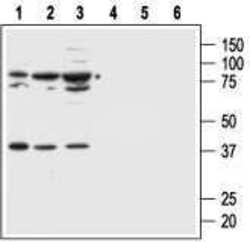
- Western blot analysis of human normal skin fibroblast cell line Malme-3 (lanes 1 and 4) and human malignant melanoma cell lines Malme-3M (lanes 2 and 5) and A875 (lanes 3 and 6): - 1,2,3. Anti-MC1 Receptor Antibody (#AMR-020), (1:500).4,5,6. Anti-MC1 Receptor Antibody , preincubated with MC1 Receptor Blocking Peptide (#BLP-MR020).
- Submitted by
- Invitrogen Antibodies (provider)
- Main image
- Experimental details
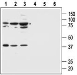
- Western blot analysis of human normal skin fibroblast cell line Malme-3 (lanes 1 and 4) and human malignant melanoma cell lines Malme-3M (lanes 2 and 5) and A875 (lanes 3 and 6): - 1,2,3. Anti-MC1 Receptor Antibody (#AMR-020), (1:500).4,5,6. Anti-MC1 Receptor Antibody , preincubated with MC1 Receptor Blocking Peptide (#BLP-MR020).

- Submitted by
- Invitrogen Antibodies (provider)
- Main image

- Experimental details
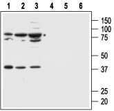
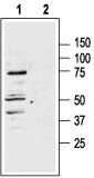
- Western blot analysis of rat adrenal lysate: - 1. Anti-MC1 Receptor Antibody (#AMR-020), (1:400). 2. Anti-MC1 Receptor Antibody , preincubated with MC1 Receptor Blocking Peptide (#BLP-MR020).
Supportive validation
- Submitted by
- Invitrogen Antibodies (provider)
- Main image
- Experimental details
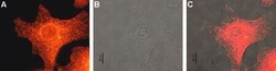
- Expression of MC1R in human Malme-3M cells - Immunocytochemical staining of human paraformaldehyde fixed and permeabilized malignant melanoma cell lines (Malme-3M). A. Cells were stained with Anti-MC1 Receptor Antibody (#AMR-020), (1:200) followed by goat- Anti-rabbit-AlexaFluor-555 secondary Antibody . B. Live view of the same field as in (A).C. Computer merge of (A) and (B).

Supportive validation
- Submitted by
- Invitrogen Antibodies (provider)
- Main image

- Experimental details
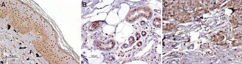
- Expressionof MC1R in normal skin and melanoma - Immunohistochemical staining of paraffin embeddednormal skin and melanoma sections using Anti-MC1 Receptor Antibody (#AMR-020) (1:100).MCR1 staining (red-brown color is highly specificin A. epidermal cells, B. eccrine sweat gland cells and C. melanoma cells. Color reaction was obtained with DAB. Hematoxilin is used as the counterstain.