Explore
 Validate
 Learn
LearnPA5-18205
antibody from Invitrogen Antibodies
Targeting: ATP6AP2
APT6M8-9, ATP6IP2, ATP6M8-9, M8-9, PRR, RENR
 Western blot
Antibody data
- Antibody Data
- Antigen structure
- References [0]
- Comments [0]
- Validations
- Western blot [4]
- Flow cytometry [2]
Submit
Validation data
Reference
Comment
Report error
- Product number
- PA5-18205 - Provider product page

- Provider
- Invitrogen Antibodies
- Product name
- ATP6IP2 Polyclonal Antibody
- Antibody type
- Polyclonal
- Antigen
- Synthetic peptide
- Description
- This antibody is predicted to react with rat based on sequence homology. This antibody is tested in Peptide ELISA: antibody detection limit dilution 8,000.
- Reactivity
- Human, Rat
- Host
- Goat
- Isotype
- IgG
- Vial size
- 100 μg
- Concentration
- 0.5 mg/mL
- Storage
- -20°C, Avoid Freeze/Thaw Cycles
No comments: Submit comment
Supportive validation
- Submitted by
- Invitrogen Antibodies (provider)
- Main image
- Experimental details
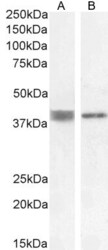
- Western blot analysis of ATP6IP2 using ATP6IP2 Polyclonal Antibody (Product # PA5-18205) (0.5 µg/mL) in staining of Human Cerebellum (A) and Heart (B) lysate (35 µg protein in RIPA buffer). Detected by chemiluminescence.

- Submitted by
- Invitrogen Antibodies (provider)
- Main image
- Experimental details
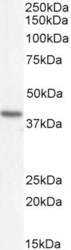
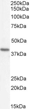
- Western blot analysis of ATP6IP2 using ATP6IP2 Polyclonal Antibody (Product # PA5-18205) (0.5 µg/mL) in staining of Rat Heart lysate (35 µg protein in RIPA buffer). Detected by chemiluminescence.
- Submitted by
- Invitrogen Antibodies (provider)
- Main image

- Experimental details
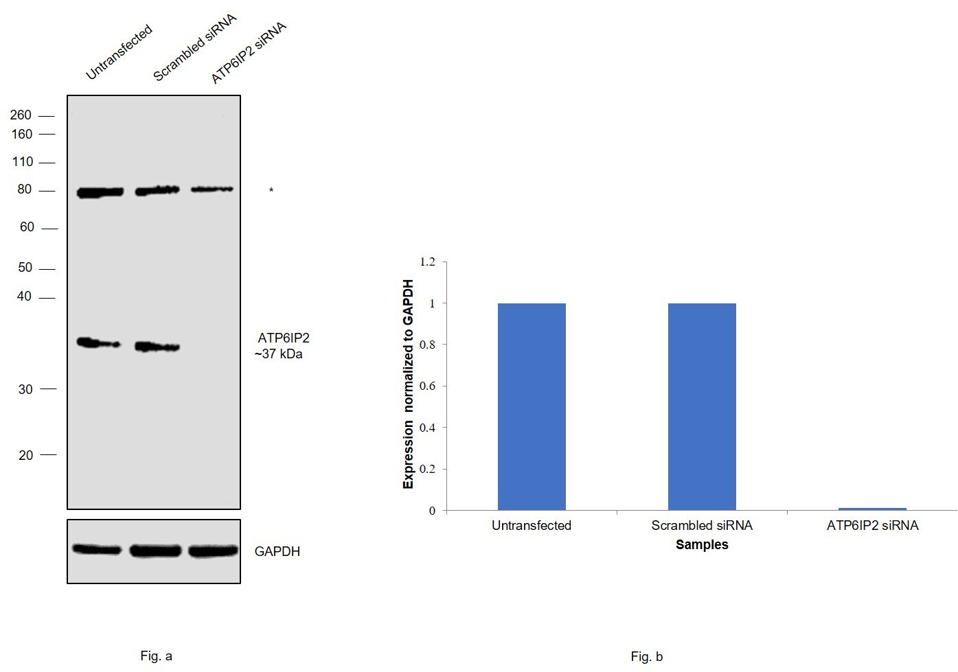
- Knockdown of ATP6IP2 was achieved by transfecting MCF-7 with ATP6IP2 specific siRNAs (Silencer® select Product # s19791, s19790). Western blot analysis (Fig. a) was performed using whole cell extracts from the ATP6IP2 knockdown cells (lane 3), non-specific scrambled siRNA transfected cells (lane 2) and untransfected cells (lane 1). The blot was probed with Arp3 Polyclonal Antibody (Product # PA5-18205, 2 µg/mL) and Rabbit anti-Goat IgG Heavy Chain Superclonal™ Recombinant Secondary Antibody, HRP (Product # A27014, 1:4,000 dilution). Densitometric analysis of this western blot is shown in histogram (Fig. b). Decrease in signal upon siRNA mediated knock down confirms that antibody is specific to ATP6IP2.
- Submitted by
- Invitrogen Antibodies (provider)
- Main image
- Experimental details
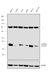
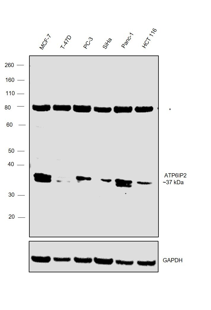
- Western blot was performed using Anti-ATP6IP2 Polyclonal Antibody (Product # PA5-18205) and a ~37 kD band was observed across the panel tested. An uncharacterized band (*) at ~80 kDa was also detected in in all the cell lines. Whole cell extracts (30 µg lysate) of MCF-7 (Lane 1), T-47D (Lane 2), PC-3 (Lane 3), SiHa (Lane 4), Panc-1 (Lane 5) and HCT 116 (Lane 6) were electrophoresed using NuPAGE™ 10% Bis-Tris Protein Gel (Product # NP0302BOX). Resolved proteins were then transferred onto a nitrocellulose membrane (Product # IB23001) by iBlot® 2 Dry Blotting System (Product # IB21001). The blot was probed with the primary antibody (2 µg/mL) and detected by chemiluminescence with Rabbit anti-Goat IgG Heavy Chain Superclonal™ Recombinant Secondary Antibody, HRP (Product # A27014, 1:4,000 dilution) using the iBright FL 1000 (Product # A32752). Chemiluminescent detection was performed using Novex® ECL Chemiluminescent Substrate Reagent Kit (Product # WP20005).
Supportive validation
- Submitted by
- Invitrogen Antibodies (provider)
- Main image
- Experimental details
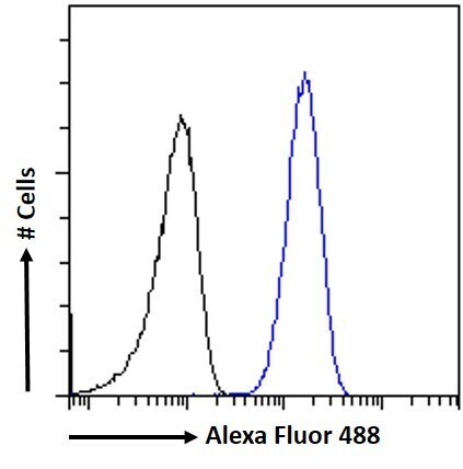
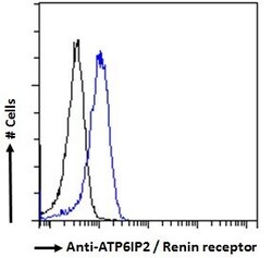
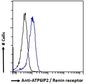
- Flow cytometric analysis of ATP6IP2 in HeLa cells using a polyclonal antibody (Product #PA5-18205). HeLa cells (blue line) were paraformaldehyde fixed and permeabilized with 0.5% Triton. The primary antibody was incubated for one hour (10 µg/mL) followed by an Alexa Fluor 488 secondary antibody (2 µg/mL). IgG control: Unimmunized goat IgG (black line) followed by an Alexa Fluor 488 secondary antibody.
- Submitted by
- Invitrogen Antibodies (provider)
- Main image
- Experimental details
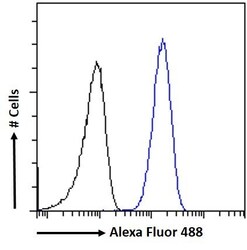
- Flow Cytometry analysis of ATP6IP2 using ATP6IP2 Polyclonal Antibody (Product # PA5-18205) in paraformaldehyde fixed HeLa cells (blue line), permeabilized with 0.5% Triton. Primary incubation 1hr (10 µg/mL) followed by Alexa Fluor 488 secondary antibody (2 µg/mL). IgG control: Unimmunized goat IgG (black line) followed by Alexa Fluor 488 secondary antibody.