Explore
 Validate
 Learn
Learn Western blot
 Immunohistochemistry
Antibody data
- Antibody Data
- Antigen structure
- References [2]
- Comments [0]
- Validations
- Immunohistochemistry [1]
Submit
Validation data
Reference
Comment
Report error
- Product number
- PB9603 - Provider product page

- Provider
- Boster Biological Technology
- Product name
- Anti-Indoleamine 2, 3-dioxygenase/IDO1 Antibody Picoband™
- Antibody type
- Polyclonal
- Description
- Polyclonal antibody for IDO/IDO1 detection. Host: Rabbit.Size: 100μg/vial. Tested applications: WB, IHC-P, ICC/IF, FCM. Reactive species: Human. IDO/IDO1 information: Molecular Weight: 45326 MW; .
- Reactivity
- Human
- Host
- Rabbit
- Vial size
- 100μg/vial
- Concentration
- Add 0.2ml of distilled water will yield a concentration of 500ug/ml.
- Storage
- At -20°C for one year. After reconstitution, at 4°C for one month. It can also be aliquoted and stored frozen at -20°C for a longer time. Avoid repeated freezing and thawing.
- Handling
- Add 0.2ml of distilled water will yield a concentration of 500ug/ml.
Submitted references Deciphering the tumor immune microenvironment of imatinib-resistance in advanced gastrointestinal stromal tumors at single-cell resolution.
Single-cell RNA sequencing reveals the cellular and molecular characteristics of high-grade and metastatic bladder cancer.
Liu X, Yu J, Li Y, Shi H, Jiao X, Liu X, Guo D, Li Z, Tian Y, Dai F, Niu Z, Zhou Y
Cell death & disease 2024 Mar 5;15(3):190
Cell death & disease 2024 Mar 5;15(3):190
Single-cell RNA sequencing reveals the cellular and molecular characteristics of high-grade and metastatic bladder cancer.
Zheng Y, Wang X, Yang X, Xing N
Cellular oncology (Dordrecht, Netherlands) 2023 Oct;46(5):1415-1427
Cellular oncology (Dordrecht, Netherlands) 2023 Oct;46(5):1415-1427
No comments: Submit comment
Supportive validation
- Submitted by
- Boster Biological Technology (provider)
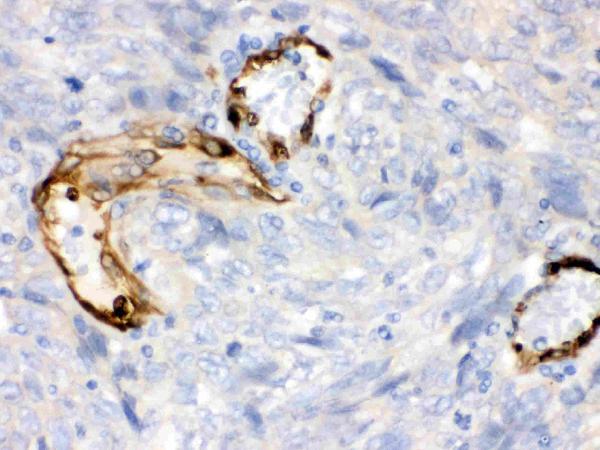
- Main image
- Experimental details
- IHC analysis of IDO1 using anti-IDO1 antibody (PB9603). IDO1 was detected in paraffin-embedded section of Human Lung Cancer Tissue. Heat mediated antigen retrieval was performed in citrate buffer (pH6, epitope retrieval solution) for 20 mins. The tissue section was blocked with 10% goat serum. The tissue section was then incubated with 1μg/ml rabbit anti-IDO1 Antibody (PB9603) overnight at 4°C. Biotinylated goat anti-rabbit IgG was used as secondary antibody and incubated for 30 minutes at 37°C. The tissue section was developed using Strepavidin-Biotin-Complex (SABC)(Catalog # SA1022) with DAB as the chromogen.
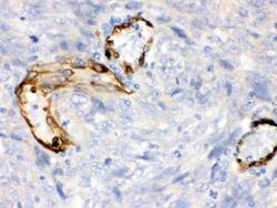
- Additional image