Explore
 Validate
 Learn
Learn Flow cytometry
Antibody data
- Antibody Data
- Antigen structure
- References [7]
- Comments [0]
- Validations
- Flow cytometry [1]
- Other assay [1]
Submit
Validation data
Reference
Comment
Report error
- Product number
- 12-9477-41 - Provider product page

- Provider
- Invitrogen Antibodies
- Product name
- IDO Monoclonal Antibody (eyedio), PE, eBioscience™
- Antibody type
- Monoclonal
- Antigen
- Other
- Description
- Description: This eyedio monoclonal antibody reacts with human indoleamine-2,3-dioxygenase (IDO, INDO, IDO1), an intracellular enzyme that catalyzes the degradation of tryptophan to kynurenines. IDO is expressed in a wide variety of tissues and cells, including macrophages, plasmacytoid dendritic cells, and several cell lines. IDO can be induced in many different cell types by IFN gamma or other inflammatory stimuli. Expression of IDO in antigen presenting cells and tumors is thought to mediate immune suppression through depletion of this essential amino acid and/or by the creation of tryptophan metabolites that causeapoptosis of T cells and induction of regulatory T cells. Applications Reported: This eyedio antibody has been reported for use in intracellular staining followed by flow cytometric analysis. Applications Tested: This eyedio antibody has been pre-titrated and tested by intracellular staining and flow cytometric analysis of normal human peripheral blood cells using the Intracellular Fixation & Permeabilization Buffer Set (Product # 88-8824-00) and protocol. Please refer to Best Protocols: Protocol A: Two step protocol for (cytoplasmic) intracellular proteins. This can be used at 5 µL (0.06 µg) per test. A test is defined as the amount (µg) of antibody that will stain a cell sample in a final volume of 100 µL. Cell number should be determined empirically but can range from 10^5 to 10^8 cells/test. Excitation: 488-561 nm; Emission: 578 nm; Laser: Blue Laser, Green Laser, Yellow-Green Laser. Filtration: 0.2 µm post-manufacturing filtered.
- Reactivity
- Human
- Host
- Mouse
- Conjugate
- Yellow dye
- Isotype
- IgG
- Antibody clone number
- eyedio
- Vial size
- 25 Tests
- Concentration
- 5 µL/Test
- Storage
- 4° C, store in dark, DO NOT FREEZE!
Submitted references IL-10(-/-) Enhances DCs Immunity Against Chlamydia psittaci Infection via OX40L/NLRP3 and IDO/Treg Pathways.
Development of a human umbilical cord-derived mesenchymal stromal cell-based advanced therapy medicinal product to treat immune and/or inflammatory diseases.
Continuing Effect of Cytokines and Toll-Like Receptor Agonists on Indoleamine-2,3-Dioxygenase-1 in Human Periodontal Ligament Stem/Stromal Cells.
HLA-DR in Cytotoxic T Lymphocytes Predicts Breast Cancer Patients' Response to Neoadjuvant Chemotherapy.
Immune Checkpoint Inhibition Overcomes ADCP-Induced Immunosuppression by Macrophages.
Clinical significance of tryptophan catabolism in Hodgkin lymphoma.
Brugia malayi microfilariae induce a regulatory monocyte/macrophage phenotype that suppresses innate and adaptive immune responses.
Li Q, Li X, Quan H, Wang Y, Qu G, Shen Z, He C
Frontiers in immunology 2021;12:645653
Frontiers in immunology 2021;12:645653
Development of a human umbilical cord-derived mesenchymal stromal cell-based advanced therapy medicinal product to treat immune and/or inflammatory diseases.
Mebarki M, Iglicki N, Marigny C, Abadie C, Nicolet C, Churlaud G, Maheux C, Boucher H, Monsel A, Menasché P, Larghero J, Faivre L, Cras A
Stem cell research & therapy 2021 Nov 13;12(1):571
Stem cell research & therapy 2021 Nov 13;12(1):571
Continuing Effect of Cytokines and Toll-Like Receptor Agonists on Indoleamine-2,3-Dioxygenase-1 in Human Periodontal Ligament Stem/Stromal Cells.
Behm C, Blufstein A, Gahn J, Kubin B, Moritz A, Rausch-Fan X, Andrukhov O
Cells 2020 Dec 16;9(12)
Cells 2020 Dec 16;9(12)
HLA-DR in Cytotoxic T Lymphocytes Predicts Breast Cancer Patients' Response to Neoadjuvant Chemotherapy.
Saraiva DP, Jacinto A, Borralho P, Braga S, Cabral MG
Frontiers in immunology 2018;9:2605
Frontiers in immunology 2018;9:2605
Immune Checkpoint Inhibition Overcomes ADCP-Induced Immunosuppression by Macrophages.
Su S, Zhao J, Xing Y, Zhang X, Liu J, Ouyang Q, Chen J, Su F, Liu Q, Song E
Cell 2018 Oct 4;175(2):442-457.e23
Cell 2018 Oct 4;175(2):442-457.e23
Clinical significance of tryptophan catabolism in Hodgkin lymphoma.
Masaki A, Ishida T, Maeda Y, Ito A, Suzuki S, Narita T, Kinoshita S, Takino H, Yoshida T, Ri M, Kusumoto S, Komatsu H, Inagaki H, Ueda R, Choi I, Suehiro Y, Iida S
Cancer science 2018 Jan;109(1):74-83
Cancer science 2018 Jan;109(1):74-83
Brugia malayi microfilariae induce a regulatory monocyte/macrophage phenotype that suppresses innate and adaptive immune responses.
O'Regan NL, Steinfelder S, Venugopal G, Rao GB, Lucius R, Srikantam A, Hartmann S
PLoS neglected tropical diseases 2014 Oct;8(10):e3206
PLoS neglected tropical diseases 2014 Oct;8(10):e3206
No comments: Submit comment
Supportive validation
- Submitted by
- Invitrogen Antibodies (provider)
- Main image
- Experimental details
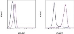
- LEFT: Surface staining of normal human peripheral blood cells with Anti-Human CD3 PerCP-eFluor® 710 (Product # 46-0036-42), Anti-Human CD11c FITC (Product # 11-0116-42), Anti-Human CD85g (ILT7) APC (Product # 17-5179-42), and Anti-Human CD123 eFluor® 450 (Product # 48-1239-42) followed by staining with Fixable Viability Dye eFluor® 506 (Product # 65-0866-14). The cells were then intracellularly stained with Anti-Human IDO PE, using the Intracellular Fixation & Permeabilization Buffer Set (Product # 88-8824-00) and protocol. Single, viable cells in the CD3+CD11c. (blue histogram) and CD85g+CD123+ (purple histogram) gates were used for analysis. RIGHT: Surface staining of unstimulated (blue histogram) or overnight LPS-stimulated (purple histogram) normal human peripheral blood cells with Anti-Human CD3 PerCP-eFluor® 710 and Anti-Human CD11c FITC followed by staining with Fixable Viability Dye eFluor® 506. The cells were then intracellularly stained with Anti-Human IDO PE, using the Intracellular Fixation & Permeabilization Buffer Set and protocol. Single, viable monocytes in the CD3-CD11c+ gate were used for analysis.
- Conjugate
- Yellow dye

Supportive validation
- Submitted by
- Invitrogen Antibodies (provider)
- Main image
- Experimental details
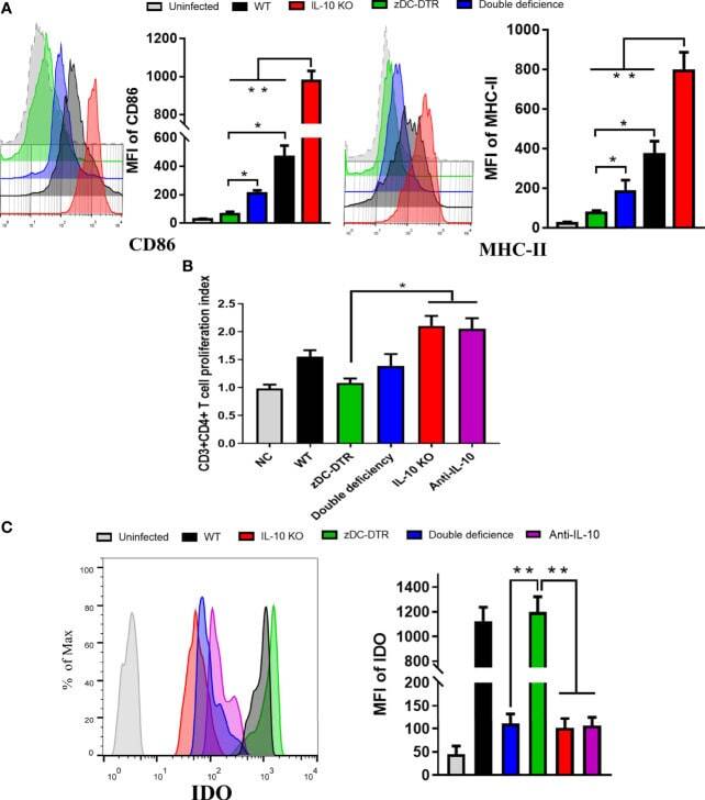
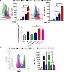
- Figure 5 DCs maturation, T cell proliferation, and IDO expression in the co-culture system. (A) Flow cytometry detection of CD86 and MHC-II DC surface markers; mean fluorescence intensity (MFI) was calculated at 72 hpi. CD86 and MHC-II were analyzed by pre-gated CD11c + cells, CD11c was gated by isotype. CD86 and MHC-II surface markers were significantly upregulated in the co-culture system of CD4 + T cells and IL-10 -/- DCs. Compared to the DCs from the zDC-DTR group, CD86 and MHC-II were significantly higher in the DD group. (B) Measurement of CD4 + T cell proliferation via BrdU assay in the co-culture system of CD4 + T cells and DCs at 72 h. Higher CD4 + T cell proliferation was observed in the IL-10 -/- and anti-IL-10 groups than the zDC-DTR group ( P
- Conjugate
- Yellow dye