 Explore
 Validate
 Learn
Learn Western blot
Antibody data
- Antibody Data
- Antigen structure
- References [6]
- Comments [0]
- Validations
- Western blot [2]
- Immunohistochemistry [1]
Submit
Validation data
Reference
Comment
Report error
- Product number
- 14-9750-82 - Provider product page

- Provider
- Invitrogen Antibodies
- Product name
- IDO Monoclonal Antibody (V1NC3IDO), eBioscience™
- Antibody type
- Monoclonal
- Antigen
- Other
- Description
- Description: The monoclonal antibody V1NC3IDO recognizes human IDO. IDO (indoleamine-2,3-dioxygenase, INDO, IDO1) is an intracellular enzyme that catalyzes the degradation of tryptophan to kynurenines. IDO is expressed in a wide variety of tissues and cells, including macrophages, plasmacytoid dendritic cells, and several cell lines. IDO can be induced in many different cell types by IFN gamma or other inflammatory stimuli. Expression of IDO in antigen presenting cells and tumors is thought to mediate immune suppression through depletion of this essential amino acid and/or by the creation of tryptophan metabolites that cause apoptosis of T cells and induction of regulatory T cells. This V1NC3IDO antibody reacts with IDO1 and does not cross react with IDO2. Applications Reported: This V1NC3IDO antibody has been reported for use in immunohistochemical staining of formalin-fixed paraffin embedded tissue sections, and microscopy. Applications Tested: This V1NC3IDO antibody has been tested by immunohistochemistry of formalin-fixed paraffin embedded human tissue using low pH antigen retrieval and can be used at less than or equal to 0.5 µg/mL. It is recommended that the antibody be carefully titrated for optimal performance in the assay of interest. Purity: Greater than 90%, as determined by SDS-PAGE. Aggregation: Less than 10%, as determined by HPLC. Filtration: 0.2 µm post-manufacturing filtered.
- Reactivity
- Human
- Host
- Mouse
- Isotype
- IgG
- Antibody clone number
- V1NC3IDO
- Vial size
- 100 µg
- Concentration
- 0.5 mg/mL
- Storage
- 4° C
Submitted references A phase 1/2 trial of an immune-modulatory vaccine against IDO/PD-L1 in combination with nivolumab in metastatic melanoma.
Th17 immune microenvironment in Epstein-Barr virus-negative Hodgkin lymphoma: implications for immunotherapy.
The role of indoleamine 2,3-dioxygenase in the induction of immune tolerance: focus on hematology.
The indoleamine 2,3-dioxygenase pathway is essential for human plasmacytoid dendritic cell-induced adaptive T regulatory cell generation.
Expression of indoleamine 2, 3-dioxygenase and the recruitment of Foxp3-expressing regulatory T cells in the development and progression of uterine cervical cancer.
Mechanism of interferon-gamma action. Characterization of indoleamine 2,3-dioxygenase in cultured human cells induced by interferon-gamma and evaluation of the enzyme-mediated tryptophan degradation in its anticellular activity.
Kjeldsen JW, Lorentzen CL, Martinenaite E, Ellebaek E, Donia M, Holmstroem RB, Klausen TW, Madsen CO, Ahmed SM, Weis-Banke SE, Holmström MO, Hendel HW, Ehrnrooth E, Zocca MB, Pedersen AW, Andersen MH, Svane IM
Nature medicine 2021 Dec;27(12):2212-2223
Nature medicine 2021 Dec;27(12):2212-2223
Th17 immune microenvironment in Epstein-Barr virus-negative Hodgkin lymphoma: implications for immunotherapy.
Duffield AS, Ascierto ML, Anders RA, Taube JM, Meeker AK, Chen S, McMiller TL, Phillips NA, Xu H, Ogurtsova A, Berger AE, Pardoll DM, Topalian SL, Ambinder RF
Blood advances 2017 Jul 25;1(17):1324-1334
Blood advances 2017 Jul 25;1(17):1324-1334
The role of indoleamine 2,3-dioxygenase in the induction of immune tolerance: focus on hematology.
Curti A, Trabanelli S, Salvestrini V, Baccarani M, Lemoli RM
Blood 2009 Mar 12;113(11):2394-401
Blood 2009 Mar 12;113(11):2394-401
The indoleamine 2,3-dioxygenase pathway is essential for human plasmacytoid dendritic cell-induced adaptive T regulatory cell generation.
Chen W, Liang X, Peterson AJ, Munn DH, Blazar BR
Journal of immunology (Baltimore, Md. : 1950) 2008 Oct 15;181(8):5396-404
Journal of immunology (Baltimore, Md. : 1950) 2008 Oct 15;181(8):5396-404
Expression of indoleamine 2, 3-dioxygenase and the recruitment of Foxp3-expressing regulatory T cells in the development and progression of uterine cervical cancer.
Nakamura T, Shima T, Saeki A, Hidaka T, Nakashima A, Takikawa O, Saito S
Cancer science 2007 Jun;98(6):874-81
Cancer science 2007 Jun;98(6):874-81
Mechanism of interferon-gamma action. Characterization of indoleamine 2,3-dioxygenase in cultured human cells induced by interferon-gamma and evaluation of the enzyme-mediated tryptophan degradation in its anticellular activity.
Takikawa O, Kuroiwa T, Yamazaki F, Kido R
The Journal of biological chemistry 1988 Feb 5;263(4):2041-8
The Journal of biological chemistry 1988 Feb 5;263(4):2041-8
No comments: Submit comment
Supportive validation
- Submitted by
- Invitrogen Antibodies (provider)
- Main image

- Experimental details
- Knockout of IDO1 was achieved by CRISPR-Cas9 genome editing using LentiArray™ Lentiviral sgRNA (Product # A32042, Assay ID CRISPR864924_LV) and LentiArray Cas9 Lentivirus (Product # A32064). Western blot analysis of IDO1 was performed by loading 30 µg of HeLa wild type (Lane 1), HeLa wild type treated with 50 ng/mL IFN gamma for 24hrs (Lane 2), HeLa Cas9 (Lane 3), HeLa Cas9 treated with 50 ng/mL IFN gamma for 24hrs (Lane 4), HeLa IDO1 KO (Lane 5) and HeLa IDO1 KO treated with 50 ng/mL IFN gamma for 24hrs (Lane 6) whole cell extracts. The samples were electrophoresed using NuPAGE™ Novex™ 4-12% Bis-Tris Protein Gel (Product # NP0322BOX). Resolved proteins were then transferred onto a nitrocellulose membrane (Product # IB23001) by iBlot® 2 Dry Blotting System (Product # IB21001). The blot was probed with Anti-IDO Monoclonal Antibody (V1NC3IDO), eBioscience™ (Product # 14-9750-82, 1;1000 dilution) and Goat anti-Mouse IgG (H+L) Superclonal™ Recombinant Secondary Antibody, HRP (Product # A28177, 1;8000 dilution) using the iBright FL 1000 (Product # A32752). Chemiluminescent detection was performed using Novex® ECL Chemiluminescent Substrate Reagent Kit (Product # WP20005). Loss of signal upon CRISPR mediated knockout (KO) using the LentiArray™ CRISPR product line confirms that antibody is specific to IDO1.

- Submitted by
- Invitrogen Antibodies (provider)
- Main image
- Experimental details
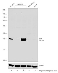
- Western blot was performed using Anti-IDO Monoclonal Antibody (V1NC3IDO), eBioscience™ (Product # 14-9750-82) and a 43 kDa band corresponding to Indoleamine 2,3-dioxygenase 1 was observed across SK-OV-3 and HEK-293 treated with IFN gamma. Whole cell extracts (30 µg lysate) of SK-O-V3 (Lane 1), HEK-293 (Lane 2), HEK-293 treated with 50 ng/ml of IFN gamma for 24 hours (Lane 3), MDA-MB-231 (Lane 4) and MCF7 (Lane 5) were electrophoresed using NuPAGE™ 4-12% Bis-Tris Protein Gel (Product # NP0321BOX). Resolved proteins were then transferred onto a Nitrocellulose membrane (Product # IB23001) by iBlot® 2 Dry Blotting System (Product # IB21001). The blot was probed with the primary antibody (1:1000 dilution) and detected by chemiluminescence with Goat anti-Mouse IgG (H+L) Superclonal™ Recombinant Secondary Antibody, HRP (Product # A28177, 1:8000 dilution) using the iBright FL 1000 (Product # A32752). Chemiluminescent detection was performed using Novex® ECL Chemiluminescent Substrate Reagent Kit (Product # WP20005).

Supportive validation
- Submitted by
- Invitrogen Antibodies (provider)
- Main image
- Experimental details
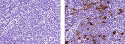
- Immunohistochemistry of formalin-fixed paraffin embedded human tonsil tissue using 0.5 µg/mL of Mouse IgG2b K Isotype Control Purified (left) or 0.5 µg/mL of Anti-Human IDO Purified (right) followed by Anti-Mouse IgG Biotin, Streptavidin-HRP, and DAB visualization.Nuclei are counterstained with hematoxylin.
