Explore
 Validate
 Learn
Learn Western blot
Antibody data
- Antibody Data
- Antigen structure
- References [0]
- Comments [0]
- Validations
- Western blot [3]
- Immunocytochemistry [2]
- Immunohistochemistry [2]
Submit
Validation data
Reference
Comment
Report error
- Product number
- PA5-12305 - Provider product page

- Provider
- Invitrogen Antibodies
- Product name
- IDO Polyclonal Antibody
- Antibody type
- Polyclonal
- Antigen
- Synthetic peptide
- Reactivity
- Human
- Host
- Rabbit
- Isotype
- IgG
- Vial size
- 400 μL
- Storage
- Store at 4°C short term. For long term storage, store at -20°C, avoiding freeze/thaw cycles.
No comments: Submit comment
Supportive validation
- Submitted by
- Invitrogen Antibodies (provider)
- Main image
- Experimental details
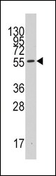
- Western blot analysis of IDO in 293 cell line lysates. Samples were incubated with IDO polyclonal antibody (Product # PA5-12305). Lysates: 35 µg/lane. INDO (arrow).
- Submitted by
- Invitrogen Antibodies (provider)
- Main image
- Experimental details
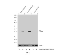
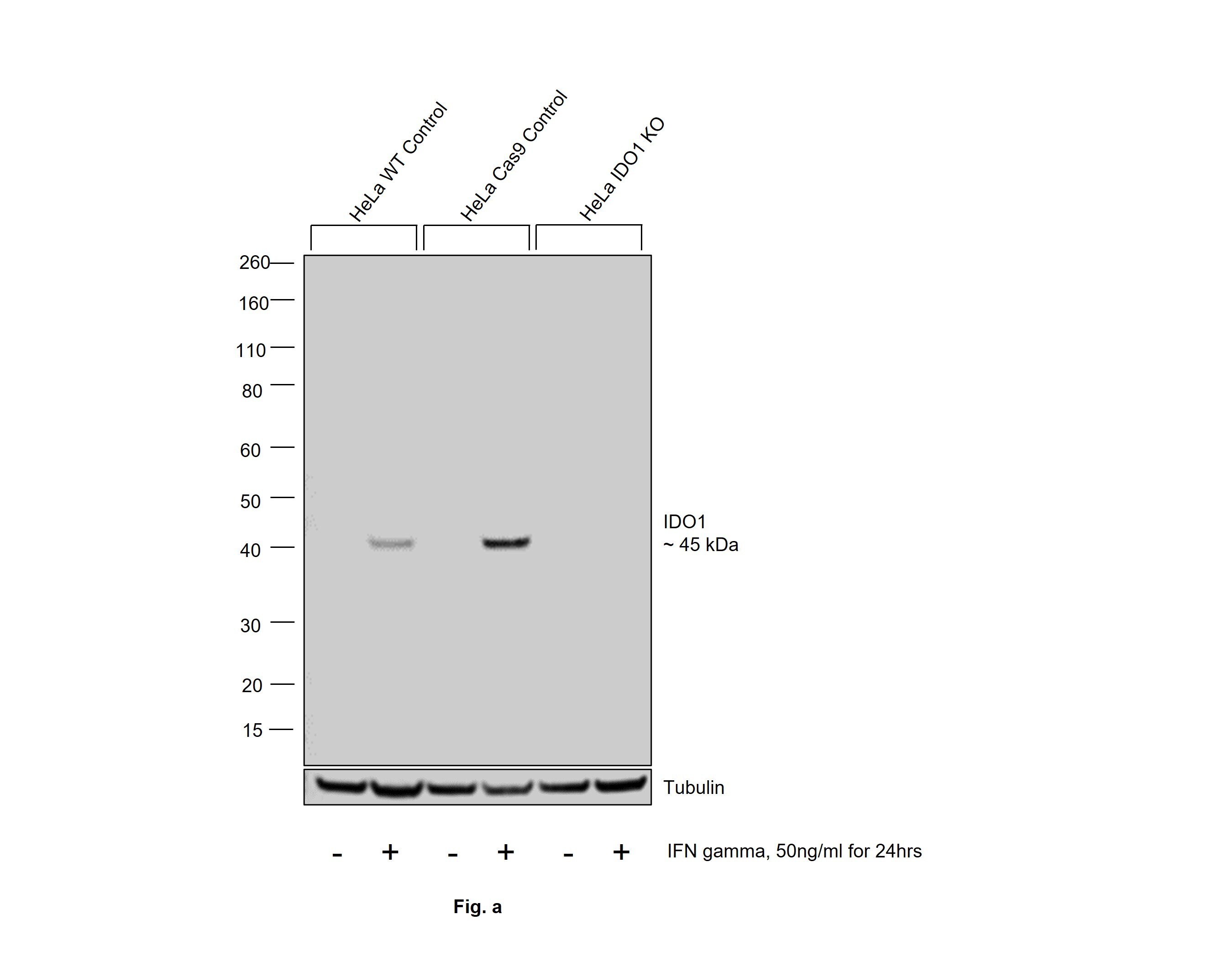
- Knockout of IDO1 was achieved by CRISPR-Cas9 genome editing using LentiArray™ Lentiviral sgRNA (Product # A32042, Assay ID CRISPR864924_LV) and LentiArray Cas9 Lentivirus (Product # A32064). Western blot analysis of IDO1 was performed by loading 40 µg of HeLa wild type (Lane 1), HeLa wild type treated with 50 ng/mL IFN gamma for 24hrs (Lane 2), HeLa Cas9 (Lane 3), HeLa Cas9 treated with 50 ng/mL IFN gamma for 24hrs (Lane 4), HeLa IDO1 KO (Lane 5) and HeLa IDO1 KO treated with 50 ng/mL IFN gamma for 24hrs (Lane 6) whole cell extracts. The samples were electrophoresed using NuPAGE™ Novex™ 4-12% Bis-Tris Protein Gel (Product # NP0322BOX). Resolved proteins were then transferred onto a nitrocellulose membrane (Product # IB23001) by iBlot® 2 Dry Blotting System (Product # IB21001). The blot was probed with Anti-IDO Polyclonal Antibody (Product # PA5-12305, 1;2000 dilution) and Goat anti-Rabbit IgG (Heavy Chain) Superclonal™ Recombinant Secondary Antibody, HRP (Product # A27036, 1;20000 dilution) using the iBright FL 1000 (Product # A32752). Chemiluminescent detection was performed using Novex® ECL Chemiluminescent Substrate Reagent Kit (Product # WP20005). Loss of signal upon CRISPR mediated knockout (KO) using the LentiArray™ CRISPR product line confirms that antibody is specific to IDO1.
- Submitted by
- Invitrogen Antibodies (provider)
- Main image
- Experimental details
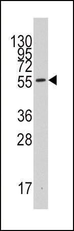
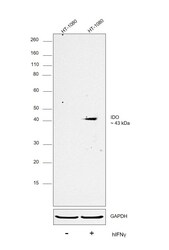
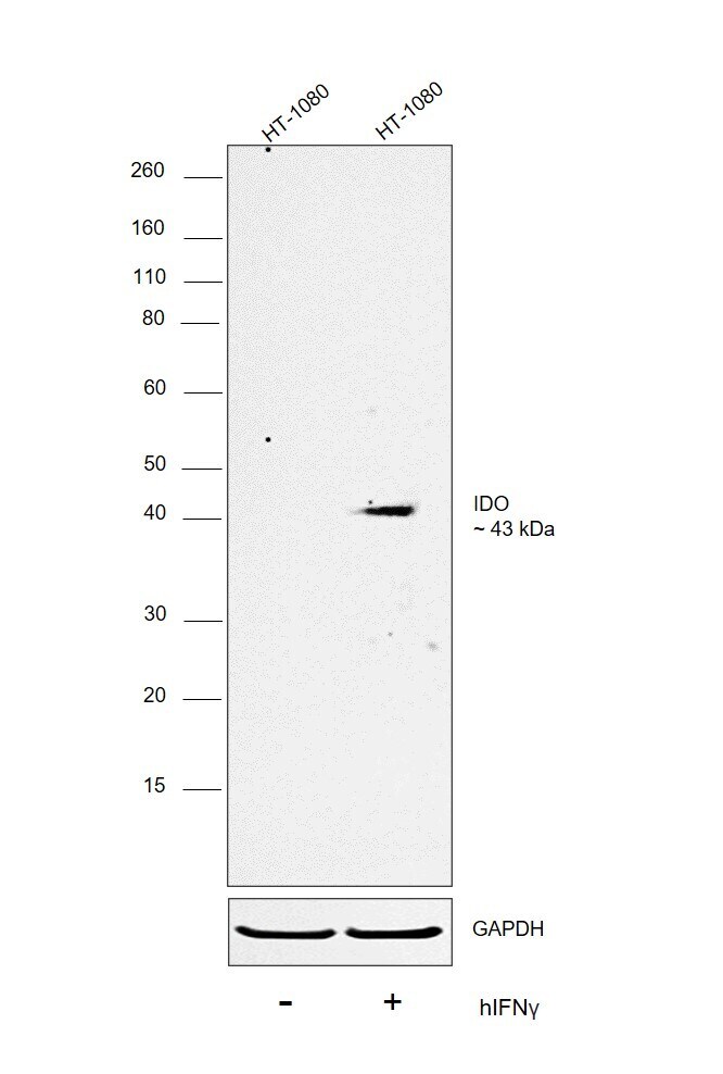
- Western blot was performed using Anti-IDO Polyclonal Antibody (Product # PA5-12305) and a 43 kDa band corresponding to Indoleamine 2,3-dioxygenase 1 was observed across cell line. Whole cell extracts (40 µg lysate) of HT-1080 untreated (Lane 1), HT-1080 treated with human interferon gamma at 50 ng/mL for 24 hrs (Lane 2) were electrophoresed using NuPAGE™ 10% Bis-Tris Protein Gel (Product # NP0301BOX). Resolved proteins were then transferred onto a nitrocellulose membrane (Product # LC2001) by iBlot® 2 Dry Blotting System (Product # IB21001). The blot was probed with the primary antibody (1:2000 dilution) and detected by chemiluminescence with Goat anti-Rabbit IgG (Heavy Chain) Superclonal™ Recombinant Secondary Antibody, HRP (Product # A27036,1:20000 dilution) using the iBright FL 1000 (Product # A32752). Chemiluminescent detection was performed using SuperSignal™ West Dura Extended Duration Substrate (Product # 34076). A clear upregulation was observed in the second lane upon treatment with human IFN gamma.
Supportive validation
- Submitted by
- Invitrogen Antibodies (provider)
- Main image
- Experimental details
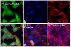
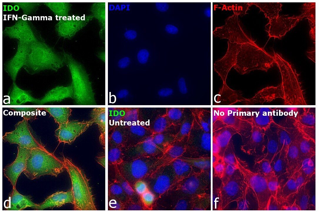
- Immunofluorescence analysis of Indoleamine 2,3-dioxygenase 1 was performed using 70% confluent log phase HT-1080 treated with human interferon gamma at 50 ng/mL concentration for 24 hrs.. The cells were fixed with 4% paraformaldehyde for 10 minutes, permeabilized with 0.1% Triton™ X-100 for 15 minutes, and blocked with 2% BSA for 1 hour at room temperature. The cells were labeled with IDO Polyclonal Antibody (Product # PA5-12305) at 1:100 dilution in 0.1% BSA, incubated at 4 degree celsius overnight and then labeled with Donkey anti-Rabbit IgG (H+L) Highly Cross-Adsorbed Secondary Antibody, Alexa Fluor Plus 488 (Product # A32790), (1:2000 dilution), for 45 minutes at room temperature (Panel a: Green). Nuclei (Panel b:Blue) were stained with ProLong™ Diamond Antifade Mountant with DAPI (Product # P36962). F-actin (Panel c: Red) was stained with Rhodamine Phalloidin (Product # R415, 1:300 dilution). Panel d represents the merged image showing cytosolic localization. Panel e represents untreated HT-1080 with very low expression. Panel f represents control cells with no primary antibody to assess background. The images were captured at 40x magnification.

- Submitted by
- Invitrogen Antibodies (provider)
- Main image

- Experimental details
- Immunofluorescence analysis of Indoleamine 2,3-dioxygenase 1 was performed using 70% confluent log phase HT-1080 treated with human interferon gamma at 50 ng/mL concentration for 24 hrs.. The cells were fixed with 4% paraformaldehyde for 10 minutes, permeabilized with 0.1% Triton™ X-100 for 15 minutes, and blocked with 2% BSA for 1 hour at room temperature. The cells were labeled with IDO Polyclonal Antibody (Product # PA5-12305) at 1:100 dilution in 0.1% BSA, incubated at 4 degree celsius overnight and then labeled with Donkey anti-Rabbit IgG (H+L) Highly Cross-Adsorbed Secondary Antibody, Alexa Fluor Plus 488 (Product # A32790), (1:2000 dilution), for 45 minutes at room temperature (Panel a: Green). Nuclei (Panel b:Blue) were stained with ProLong™ Diamond Antifade Mountant with DAPI (Product # P36962). F-actin (Panel c: Red) was stained with Rhodamine Phalloidin (Product # R415, 1:300 dilution). Panel d represents the merged image showing cytosolic localization. Panel e represents untreated HT-1080 with very low expression. Panel f represents control cells with no primary antibody to assess background. The images were captured at 40x magnification.
Supportive validation
- Submitted by
- Invitrogen Antibodies (provider)
- Main image

- Experimental details
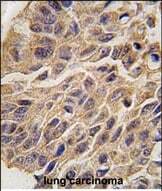
- Immunohistochemistry analysis of IDO in formalin-fixed and paraffin-embedded human lung carcinoma tissue. Samples were incubated with IDO polyclonal antibody (Product # PA5-12305) which was peroxidase-conjugated to the secondary antibody, followed by DAB staining. This data demonstrates the use of this antibody for immunohistochemistry; clinical relevance has not been evaluated.

- Submitted by
- Invitrogen Antibodies (provider)
- Main image

- Experimental details
- Immunohistochemistry analysis of IDO in formalin-fixed and paraffin-embedded human lung carcinoma tissue. Samples were incubated with IDO polyclonal antibody (Product # PA5-12305) which was peroxidase-conjugated to the secondary antibody, followed by DAB staining. This data demonstrates the use of this antibody for immunohistochemistry; clinical relevance has not been evaluated.