Explore
 Validate
 Learn
Learn Western blot
 Immunohistochemistry
Antibody data
- Antibody Data
- Antigen structure
- References [3]
- Comments [0]
- Validations
- Western blot [1]
- Immunocytochemistry [2]
- Other assay [3]
Submit
Validation data
Reference
Comment
Report error
- Product number
- MA1-80034 - Provider product page

- Provider
- Invitrogen Antibodies
- Product name
- PLP1 Monoclonal Antibody (plpc1)
- Antibody type
- Monoclonal
- Antigen
- Synthetic peptide
- Description
- Predicted to react with all mammals based on sequence homology. Mouse anti myelin proteolipid protein antibody, clone plpc1 recognizes myelin proteolipid protein (PLP) in many mammalian species (Stoffel et al. 1985).
- Reactivity
- Human, Mouse, Rat, Bovine, Canine
- Host
- Mouse
- Isotype
- IgG
- Antibody clone number
- plpc1
- Vial size
- 100 μg
- Concentration
- 1 mg/mL
- Storage
- Store at 4°C short term. For long term storage, store at -20°C, avoiding freeze/thaw cycles.
Submitted references Role of Enteric Glia as Bridging Element between Gut Inflammation and Visceral Pain Consolidation during Acute Colitis in Rats.
Plasma Exosome Profile in ST-Elevation Myocardial Infarction Patients with and without Out-of-Hospital Cardiac Arrest.
Oligodendroglial excitability mediated by glutamatergic inputs and Nav1.2 activation.
Lucarini E, Seguella L, Vincenzi M, Parisio C, Micheli L, Toti A, Corpetti C, Del Re A, Squillace S, Maftei D, Lattanzi R, Ghelardini C, Di Cesare Mannelli L, Esposito G
Biomedicines 2021 Nov 12;9(11)
Biomedicines 2021 Nov 12;9(11)
Plasma Exosome Profile in ST-Elevation Myocardial Infarction Patients with and without Out-of-Hospital Cardiac Arrest.
Zarà M, Campodonico J, Cosentino N, Biondi ML, Amadio P, Milanesi G, Assanelli E, Cerri S, Biggiogera M, Sandrini L, Tedesco CC, Veglia F, Trabattoni D, Blandini F, Tremoli E, Marenzi G, Barbieri SS
International journal of molecular sciences 2021 Jul 28;22(15)
International journal of molecular sciences 2021 Jul 28;22(15)
Oligodendroglial excitability mediated by glutamatergic inputs and Nav1.2 activation.
Berret E, Barron T, Xu J, Debner E, Kim EJ, Kim JH
Nature communications 2017 Sep 15;8(1):557
Nature communications 2017 Sep 15;8(1):557
No comments: Submit comment
Supportive validation
- Submitted by
- Invitrogen Antibodies (provider)
- Main image
- Experimental details
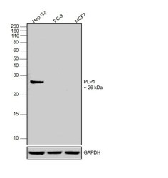
- Western blot was performed using Anti-PLP1 Monoclonal Antibody (Product # MA1-80034) and 26 kDa band corresponding to PLP1 was observed in the positive model, Hep G2 and not in negative cell lines, MCF7 and PC-3 as reported. Whole cell extracts (40 µg lysate) of Hep G2 (Lane 1), PC-3 (Lane 2) and MCF7 (Lane 3) were electrophoresed using Novex® NuPAGE® 12 % Bis-Tris gel (Product # NP0342BOX). Resolved proteins were then transferred onto a nitrocellulose membrane (Product # IB23001) by iBlot® 2 Dry Blotting System (Product # IB21001). The blot was probed with the primary antibody (1:1000 dilution) and detected by chemiluminescence with Goat anti-Mouse IgG (H+L) Superclonal™ Recombinant Secondary Antibody, HRP (Product # A28177, 1:4000 dilution) using the iBright FL 1000 (Product # A32752). Chemiluminescent detection was performed using Novex® ECL Chemiluminescent Substrate Reagent Kit (Product # WP20005)..
Supportive validation
- Submitted by
- Invitrogen Antibodies (provider)
- Main image
- Experimental details
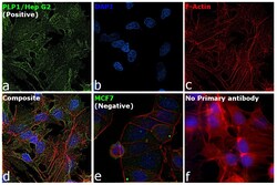
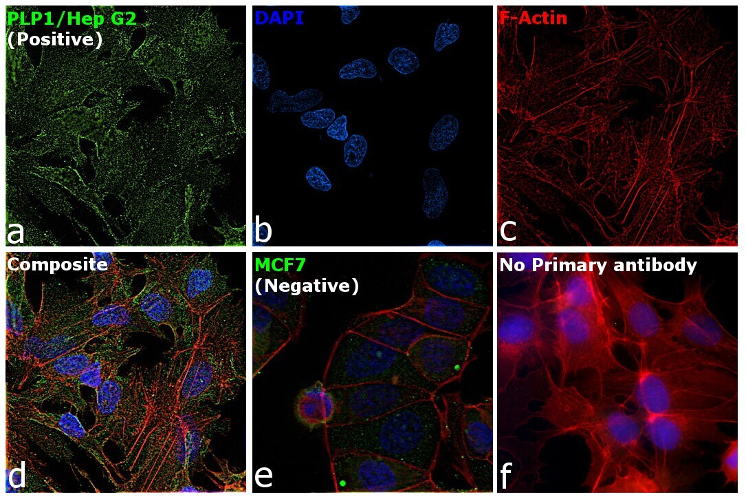
- Immunofluorescence analysis of PLP1 was performed using Hep G2 and MCF7 cells. The cells were fixed with 4% paraformaldehyde for 10 minutes, permeabilized with 0.1% Triton™ X-100 for 15 minutes, and blocked with 2% BSA for 1 hour at room temperature. The cells were labeled with PLP1 Monoclonal Antibody (plpc1) (Product # MA1-80034) at 1:100 dilution in 0.1% BSA and incubated overnight at 4 degree and then labeled with Goat anti-Mouse IgG (H+L) Highly Cross-Adsorbed Secondary Antibody, Alexa Fluor Plus 488 (Product # A32723, 1:2000 dilution) for 45 minutes at room temperature (Panel a: green). Nuclei (Panel b: blue) were stained with ProLong™ Diamond Antifade Mountant with DAPI (Product # P36962). F-actin (Panel c: red) was stained with Rhodamine Phalloidin (Product # R415, 1:300). Panel d represents the merged image showing cytoplasmic of PLP1 in Hep G2 (the positive model) and not in MCF7 (the negative model) (panel e). Panel f represents control cells with no primary antibody to assess background. The images were captured at 60X magnification.

- Submitted by
- Invitrogen Antibodies (provider)
- Main image

- Experimental details
- Immunofluorescence analysis of PLP1 was performed using Hep G2 and MCF7 cells. The cells were fixed with 4% paraformaldehyde for 10 minutes, permeabilized with 0.1% Triton™ X-100 for 15 minutes, and blocked with 2% BSA for 1 hour at room temperature. The cells were labeled with PLP1 Monoclonal Antibody (plpc1) (Product # MA1-80034) at 1:100 dilution in 0.1% BSA and incubated overnight at 4 degree and then labeled with Goat anti-Mouse IgG (H+L) Highly Cross-Adsorbed Secondary Antibody, Alexa Fluor Plus 488 (Product # A32723, 1:2000 dilution) for 45 minutes at room temperature (Panel a: green). Nuclei (Panel b: blue) were stained with ProLong™ Diamond Antifade Mountant with DAPI (Product # P36962). F-actin (Panel c: red) was stained with Rhodamine Phalloidin (Product # R415, 1:300). Panel d represents the merged image showing cytoplasmic of PLP1 in Hep G2 (the positive model) and not in MCF7 (the negative model) (panel e). Panel f represents control cells with no primary antibody to assess background. The images were captured at 60X magnification.
Supportive validation
- Submitted by
- Invitrogen Antibodies (provider)
- Main image
- Experimental details
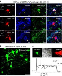
- Fig. 2 Excitable OLs belong to the pre-myelinating OL stage. a Post-recording immunostaining of Alexa 568-filled excitable OLs ( red ) for CNPase ( blue ) and DM20-PLP ( green , upper ), NG2 ( green , middle ), or MBP ( green , lower ) in the MNTB (P9-P14). b Confocal image of CNPase-GFP+ cells in the MNTB that were infected by adenovirus encoding pAV.ExSi-CNPase promoter-eGFP. c CNPase-GFP+ pre-OLs in the MNTB. A CNPase-GFP+ pre-OL was filled with Alexa 568 during whole-cell recording, as demonstrated in differential interference contrast (DIC) and fluorescence images. Dashed lines indicate the patch pipette. The CNPase-GFP+ pre-OL fired APs in response to step-current injections of 80 and 120 pA (300 ms). Scale bar , 10 mum
- Submitted by
- Invitrogen Antibodies (provider)
- Main image
- Experimental details
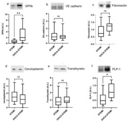
- Figure 4 Plasma exosomes of OHCA-STEMI patients display a greater expression of platelet and brain-associated markers. ( a - f ) Densitometric quantification and representative images of Western blot analysis of GPIIb ( a ), VE-cadherin ( b ), fibronectin ( c ), ceruloplasmin ( d ), transthyretin ( e ) and PLP1 ( f ). For each box plot, the center line illustrates the median and box limits indicate the 25th and 75th percentiles. ns: not significant, * p < 0.05, ** p < 0.01.

- Submitted by
- Invitrogen Antibodies (provider)
- Main image
- Experimental details
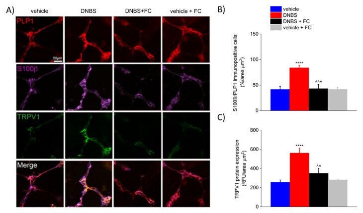
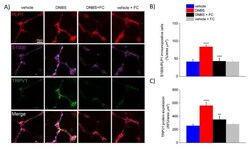
- Figure 5 Effect of systemic fluorocitrate on S100beta and TRPV1 increased expression within the colonic myenteric plexus of DNBS rats at day 7. ( A ) Immunofluorescence images show the expression of PLP1 (red), S100beta (purple), and TRPV1 (green) in the myenteric plexus of the colon and ( B , C ) relative immunolabeling quantification at day 7 after colitis induction. Data were analysed by one-way ANOVA and Bonferroni post-hoc. Results are expressed as a mean +- SEM of the percentage of PLP1 immunopositive cells that co-express S100beta per area unit (mum 2 ) of n assessments. Results about TRPV1 expression are expressed as average relative fluorescence units (RFUs) +- SEM per area unit of (mum 2 ) of n assessments. **** p < 0.0001 vs. vehicle, ^^^ p < 0.001 and ^^ p < 0.01 vs. DNBS. Original magnification: 20x. Scale bar: 50 mum.