Explore
 Validate
 Learn
Learn Western blot
Antibody data
- Antibody Data
- Antigen structure
- References [0]
- Comments [0]
- Validations
- Western blot [3]
- Immunohistochemistry [1]
Submit
Validation data
Reference
Comment
Report error
- Product number
- PA5-111891 - Provider product page

- Provider
- Invitrogen Antibodies
- Product name
- TBXA2R Polyclonal Antibody
- Antibody type
- Polyclonal
- Antigen
- Synthetic peptide
- Reactivity
- Human, Mouse, Rat
- Host
- Rabbit
- Isotype
- IgG
- Vial size
- 50 µL
- Concentration
- 0.8 mg/mL
- Storage
- -20°C
No comments: Submit comment
Supportive validation
- Submitted by
- Invitrogen Antibodies (provider)
- Main image

- Experimental details
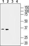
- Western Blot analysis of TBXA2R was performed in human THP-1 monocytic leukemia (lanes 1 and 3) and human MEG-01 megakaryoblastic leukemia (lanes 2 and 4) cell line lysates. Lane 1,2: TBXA2R Antibody (Product # PA5-111891) at a dilution of 1:200. Lane 3,4: TBXA2R Antibody preincubated with the negative control antigen.

- Submitted by
- Invitrogen Antibodies (provider)
- Main image

- Experimental details
- Western Blot analysis of TBXA2R was performed in human THP-1 monocytic leukemia (lanes 1 and 3) and human MEG-01 megakaryoblastic leukemia (lanes 2 and 4) cell line lysates. Lane 1,2: TBXA2R Antibody (Product # PA5-111891) at a dilution of 1:200. Lane 3,4: TBXA2R Antibody preincubated with the negative control antigen.
- Submitted by
- Invitrogen Antibodies (provider)
- Main image

- Experimental details
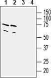
- Western Blot analysis of TBXA2R was performed in mouse (lanes 1 and 3) and rat (lanes 2 and 4) brain lysates. Lane 1,2: TBXA2R Antibody (Product # PA5-111891) at a dilution of 1:200. Lane 3,4: TBXA2R Antibody preincubated with the negative control antigen.
Supportive validation
- Submitted by
- Invitrogen Antibodies (provider)
- Main image
- Experimental details
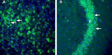
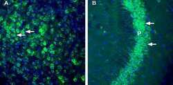
- Immunohistochemistry analysis of TBXA2R in perfusion-fixed, frozen mouse cortex and hippocampus tissue sections using TBXA2R Antibody (Product # PA5-111891) at a dilution of 1:200, followed by goat anti-rabbit-AlexaFluor-488. A) TBXA2R immunoreactivity (green) is observed in mouse parietal cortex neurons (arrows). B) TBXA2R immunoreactivity (green) is detected in neurons (horizontal arrows) of mouse hippocampal CA3 pyramidal layer (P). Cell nuclei are stained with DAPI (blue).