Explore
 Validate
 Learn
LearnPB9128
antibody from Boster Biological Technology
Targeting: APEX1
APE, APE-1, APEN, APEX, APX, HAP1, REF-1, REF1
 Western blot
 Immunocytochemistry
Antibody data
- Antibody Data
- Antigen structure
- References [1]
- Comments [0]
- Validations
- Western blot [1]
Submit
Validation data
Reference
Comment
Report error
- Product number
- PB9128 - Provider product page

- Provider
- Boster Biological Technology
- Product name
- Anti-APE1/APEX1 Antibody Picoband™
- Antibody type
- Polyclonal
- Description
- Polyclonal antibody for APE1/APEX1 detection. Host: Rabbit.Size: 100μg/vial. Tested applications: WB, IHC-P, IHC-F, ICC, IF, FCM. Reactive species: Human. APE1/APEX1 information: Molecular Weight: 35555 MW; Subcellular Localization: Nucleus. Nucleus, nucleolus. Nucleus speckle. Endoplasmic reticulum. Cytoplasm. Detected in the cytoplasm of B-cells stimulated to switch (By similarity). Colocalized with SIRT1 in the nucleus. Colocalized with YBX1 in nuclear speckles after genotoxic stress. Together with OGG1 is recruited to nuclear speckles in UVA-irradiated cells. Colocalized with nucleolin and NPM1 in the nucleolus. Its nucleolar localization is cell cycle dependent and requires active rRNA transcription. Colocalized with calreticulin in the endoplasmic reticulum. Translocation from the nucleus to the cytoplasm is stimulated in presence of nitric oxide (NO) and function in a CRM1-dependent manner, possibly as a consequence of demasking a nuclear export signal (amino acid position 64-80). S-nitrosylation at Cys-93 and Cys-310 regulates its nuclear-cytosolic shuttling. Ubiquitinated form is localized predominantly in the cytoplasm.
- Reactivity
- Human, Mouse, Rat
- Host
- Rabbit
- Vial size
- 100μg/vial
- Concentration
- Add 0.2ml of distilled water will yield a concentration of 500ug/ml.
- Storage
- At -20°C for one year. After reconstitution, at 4°C for one month. It can also be aliquoted and stored frozen at -20°C for a longer time. Avoid repeated freezing and thawing.
- Handling
- Add 0.2ml of distilled water will yield a concentration of 500ug/ml.
Submitted references Epigenetic silencing of TET1 mediated hydroxymethylation of base excision repair pathway during lung carcinogenesis.
Chen HQ, Chen DJ, Li Y, Yuan WB, Fan J, Zhang Z, Han F, Jiang X, Chen JP, Wang DD, Cao J, Liu JY, Liu WB
Environmental pollution (Barking, Essex : 1987) 2021 Jan 1;268(Pt B):115860
Environmental pollution (Barking, Essex : 1987) 2021 Jan 1;268(Pt B):115860
No comments: Submit comment
Supportive validation
- Submitted by
- Boster Biological Technology (provider)
- Main image
- Experimental details
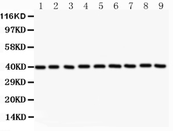
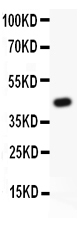
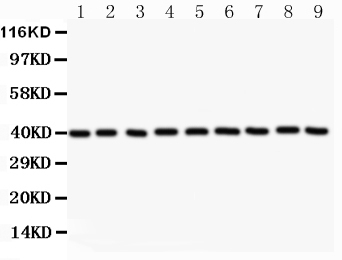
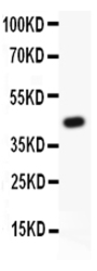
- Western blot analysis of APEX1 using anti-APEX1 antibody (PB9128). Electrophoresis was performed on a 5-20% SDS-PAGE gel at 70V (Stacking gel) / 90V (Resolving gel) for 2-3 hours. Lane 1: Recombinant Human APEX1 Protein 0.5ng. After Electrophoresis, proteins were transferred to a Nitrocellulose membrane at 150mA for 50-90 minutes. Blocked the membrane with 5% Non-fat Milk/ TBS for 1.5 hour at RT. The membrane was incubated with rabbit anti-APEX1 antigen affinity purified polyclonal antibody (Catalog # PB9128) at 0.5 μg/mL overnight at 4°C, then washed with TBS-0.1%Tween 3 times with 5 minutes each and probed with a goat anti-rabbit IgG-HRP secondary antibody at a dilution of 1:10000 for 1.5 hour at RT. The signal is developed using an Enhanced Chemiluminescent detection (ECL) kit (Catalog # EK1002) with Tanon 5200 system. A specific band was detected for APEX1 at approximately 45KD. The expected band size for APEX1 is at 45KD.
- Additional image