Explore
 Validate
 Learn
Learn Western blot
Antibody data
- Antibody Data
- Antigen structure
- References [1]
- Comments [0]
- Validations
- Western blot [2]
Submit
Validation data
Reference
Comment
Report error
- Product number
- AF1044 - Provider product page

- Provider
- R&D Systems
- Product name
- Human/Mouse/Rat APE Antibody
- Antibody type
- Polyclonal
- Description
- Antigen Affinity-purified. Detects human, mouse, and rat APE in Western blots.
- Reactivity
- Human, Mouse, Rat
- Host
- Goat
- Conjugate
- Unconjugated
- Antigen sequence
P27695- Isotype
- IgG
- Vial size
- 100 ug
- Concentration
- LYOPH
- Storage
- Use a manual defrost freezer and avoid repeated freeze-thaw cycles. 12 months from date of receipt, -20 to -70 °C as supplied. 1 month, 2 to 8 °C under sterile conditions after reconstitution. 6 months, -20 to -70 °C under sterile conditions after reconstitution.
Submitted references Differential expression of APE1 and APE2 in germinal centers promotes error-prone repair and A:T mutations during somatic hypermutation.
Stavnezer J, Linehan EK, Thompson MR, Habboub G, Ucher AJ, Kadungure T, Tsuchimoto D, Nakabeppu Y, Schrader CE
Proceedings of the National Academy of Sciences of the United States of America 2014 Jun 24;111(25):9217-22
Proceedings of the National Academy of Sciences of the United States of America 2014 Jun 24;111(25):9217-22
No comments: Submit comment
Supportive validation
- Submitted by
- R&D Systems (provider)
- Main image
- Experimental details
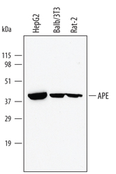
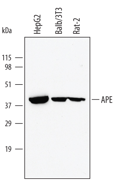
- Detection of Human, Mouse, and Rat APE by Western Blot. Western blot shows lysates of HepG2 human hepatocellular carcinoma cell line, Balb/3T3 mouse embryonic fibroblast cell line, and Rat-2 rat embryonic fibroblast cell line. PVDF membrane was probed with 1 µg/mL of Goat Anti-Human/Mouse/Rat APE Antigen Affinity-purified Polyclonal Antibody (Catalog # AF1044) followed by HRP-conjugated Anti-Goat IgG Secondary Antibody (Catalog # HAF017). A specific band was detected for APE at approximately 40 kDa (as indicated). This experiment was conducted using Immunoblot Buffer Group 1.
- Submitted by
- R&D Systems (provider)
- Main image

- Experimental details
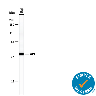
- Detection of Human APE by Simple WesternTM. Simple Western lane view shows lysates of Raji human Burkitt's lymphoma cell line, loaded at 0.2 mg/mL. A specific band was detected for APE at approximately 45 kDa (as indicated) using 10 µg/mL of Goat Anti-Human/Mouse/Rat APE Antigen Affinity-purified Polyclonal Antibody (Catalog # AF1044) followed by 1:50 dilution of HRP-conjugated Anti-Goat IgG Secondary Antibody (Catalog # HAF109). This experiment was conducted under reducing conditions and using the 12-230 kDa separation system.