Explore
 Validate
 Learn
LearnPA1665
antibody from Boster Biological Technology
Targeting: NLRP3
AGTAVPRL, AII, AVP, C1orf7, CIAS1, CLR1.1, DFNA34, FCAS, FCU, MWS, NALP3, PYPAF1
 Western blot
 Immunocytochemistry
Antibody data
- Antibody Data
- Antigen structure
- References [80]
- Comments [0]
- Validations
- Western blot [1]
Submit
Validation data
Reference
Comment
Report error
- Product number
- PA1665 - Provider product page

- Provider
- Boster Biological Technology
- Product name
- Anti-CIAS1/NALP3/NLRP3 Antibody
- Antibody type
- Polyclonal
- Description
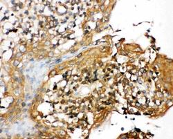
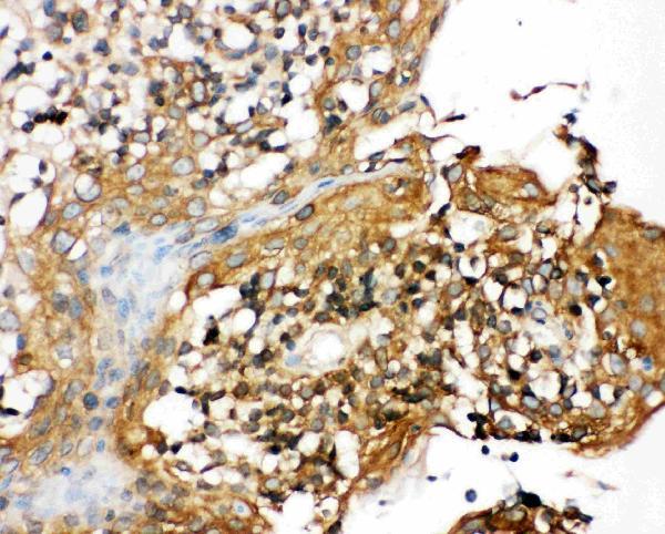
- Polyclonal antibody for NALP3/NLRP3 detection. Host: Rabbit.Size: 100μg/vial. Tested applications: IHC-P. Reactive species: Human. NALP3/NLRP3 information: Molecular Weight: 118173 MW; Subcellular Localization: Cytoplasm ; Tissue Specificity: Expressed in blood leukocytes. Strongly expressed in polymorphonuclear cells and osteoblasts. Undetectable or expressed at a lower magnitude in B- and T-lymphoblasts, respectively. High level of expression detected in chondrocytes. Detected in non-keratinizing epithelia of oropharynx, esophagus and ectocervix and in the urothelial layer of the bladder.
- Reactivity
- Human, Mouse, Rat
- Host
- Rabbit
- Vial size
- 100μg/vial
- Concentration
- Add 0.2ml of distilled water will yield a concentration of 500ug/ml.
- Storage
- At -20°C for one year. After reconstitution, at 4°C for one month. It can also be aliquoted and stored frozen at -20°C for a longer time. Avoid repeated freezing and thawing.
- Handling
- Add 0.2ml of distilled water will yield a concentration of 500ug/ml.
Submitted references Dopamine promotes Klebsiella quasivariicola proliferation and inflammatory response in the presence of macrophages.
Folic Acid Rescues Dopaminergic Neurons in MPTP-Induced Mice by Inhibiting the NLRP3 Inflammasome and Ameliorating Mitochondrial Impairment.
Phoenixin 20 ameliorates pulmonary arterial hypertension via inhibiting inflammation and oxidative stress.
PLD2 deletion ameliorates sepsis-induced cardiomyopathy by suppressing cardiomyocyte pyroptosis via the NLRP3/caspase 1/GSDMD pathway.
In vitro and in silico analyses reveal the toxicity of metolachlor to grass carp hepatocytes and the antagonism of melatonin.
Rhizoma Alismatis Decoction improved mitochondrial dysfunction to alleviate SASP by enhancing autophagy flux and apoptosis in hyperlipidemia acute pancreatitis.
ChemR23 signaling ameliorates brain injury via inhibiting NLRP3 inflammasome-mediated neuronal pyroptosis in ischemic stroke.
Dietary Cannabidiol Activates PKA/AMPK Signaling and Attenuates Chronic Inflammation and Leaky Gut in DSS-Induced Colitis Mice.
Edaravone dexborneol alleviates cerebral ischemia-reperfusion injury through NF-κB/NLRP3 signal pathway.
Transplantation of olfactory ensheathing cells can alleviate neuroinflammatory responses in rats with trigeminal neuralgia.
Metformin inhibits activation of NLRP3 inflammasome and inflammatory response in preeclamptic rats.
Carnosine ameliorates postoperative cognitive dysfunction of aged rats by limiting astrocytes pyroptosis.
Qingda granule alleviates cerebral ischemia/reperfusion injury by inhibiting TLR4/NF-κB/NLRP3 signaling in microglia.
Neutrophil Extracellular Traps Induce Alveolar Macrophage Pyroptosis by Regulating NLRP3 Deubiquitination, Aggravating the Development of Septic Lung Injury.
Pinocembrin alleviates pyroptosis and apoptosis through ROS elimination in random skin flaps via activation of SIRT3.
PM(2.5) induces the inflammatory response in rat spleen lymphocytes through autophagy activation of NLRP3 inflammasome.
Melatonin modulates the aggravation of pyroptosis, necroptosis, and neuroinflammation following cerebral ischemia and reperfusion injury in obese rats.
Pregnane X receptor (PXR) deficiency protects against spinal cord injury by activating NRF2/HO-1 pathway.
ChemR23 activation attenuates cognitive impairment in chronic cerebral hypoperfusion by inhibiting NLRP3 inflammasome-induced neuronal pyroptosis.
Heat-killed Lactobacillus murinus confers neuroprotection against dopamine neuronal loss by targeting NLRP3 inflammasome.
The capsaicinoid nonivamide suppresses the inflammatory response and attenuates the progression of steatosis in a NAFLD-rat model.
Investigations into ferroptosis in methylmercury-induced acute kidney injury in mice.
Melatonin suppresses inflammation and blood‒brain barrier disruption in rats with vascular dementia possibly by activating the SIRT1/PGC-1α/PPARγ signaling pathway.
The potential effects of festidinol treatment against the NLRP3 inflammasome and pyroptosis in D-galactose and aluminum chloride-induced Alzheimer's-like pathology in mouse brain.
Mitophagy-promoting miR-138-5p promoter demethylation inhibits pyroptosis in sepsis-associated acute lung injury.
Upregulation of TXNIP contributes to granulosa cell dysfunction in polycystic ovary syndrome via activation of the NLRP3 inflammasome.
Manf Enhances the Pyroptosis Inhibition of Bone Marrow-derived Mesenchymal Stem Cells to Relieve Cerebral Infarction Injury.
Brevilin A ameliorates sepsis-induced cardiomyopathy through inhibiting NLRP3 inflammation.
Menstrual blood-derived endometrial stem cells inhibit neuroinflammation by regulating microglia through the TLR4/MyD88/NLRP3/Casp1 pathway.
Effects of Hypoxic Environment on Periodontal Tissue through the ROS/TXNIP/NLRP3 Inflammasome Pathway.
The Hypolipidemic Effect of Hawthorn Leaf Flavonoids through Modulating Lipid Metabolism and Gut Microbiota in Hyperlipidemic Rats.
The potential mechanism of huazhuojiedu decoction in the treatment of ulcerative colitis based on network pharmacology and experimental validation.
Chronic high-fat diet consumption exacerbates pyroptosis- and necroptosis-mediated HMGB1 signaling in the brain after ischemia and reperfusion injury.
Postcooling But Not Precooling Benefits Motor Recovery by Suppressing Cell Death After Surgical Spinal Cord Injury in Rats.
Pyrroloquinoline quinone ameliorates diabetic cardiomyopathy by inhibiting the pyroptosis signaling pathway in C57BL/6 mice and AC16 cells.
Pyrroloquinoline quinone ameliorates renal fibrosis in diabetic nephropathy by inhibiting the pyroptosis pathway in C57BL/6 mice and human kidney 2 cells.
The circadian clock protein Rev-erbα provides neuroprotection and attenuates neuroinflammation against Parkinson's disease via the microglial NLRP3 inflammasome.
Daphnes Cortex and its licorice-processed products suppress inflammation via the TLR4/NF-κB/NLRP3 signaling pathway and regulation of the metabolic profile in the treatment of rheumatoid arthritis.
Sodium Butyrate Attenuates AGEs-Induced Oxidative Stress and Inflammation by Inhibiting Autophagy and Affecting Cellular Metabolism in THP-1 Cells.
Short-Chain Fatty Acids Weaken Ox-LDL-Induced Cell Inflammatory Injury by Inhibiting the NLRP3/Caspase-1 Pathway and Affecting Cellular Metabolism in THP-1 Cells.
Epigallocatechin-3-gallate ameliorates renal endoplasmic reticulum stress-mediated inflammation in type 2 diabetic rats.
The combination of dapagliflozin and statins ameliorates renal injury through attenuating the activation of inflammasome-mediated autophagy in insulin-resistant rats.
Cordycepin alleviated metabolic inflammation in Western diet-fed mice by targeting intestinal barrier integrity and intestinal flora.
Methylation Status of the miR-141-3p Promoter Regulates miR-141-3p Expression, Inflammasome Formation, and the Invasiveness of HTR-8/SVneo Cells.
Deficiency of tenascin-C attenuated cardiac injury by inactivating TLR4/NLRP3/caspase-1 pathway after myocardial infarction.
Suppression of lncRNA NLRP3 inhibits NLRP3-triggered inflammatory responses in early acute lung injury.
LncRNA 4344 promotes NLRP3-related neuroinflammation and cognitive impairment by targeting miR-138-5p.
Isoliquiritigenin alleviates LPS/ D-GalN-induced acute liver failure by activating the PGC-1α/ Nrf2 pathway to reduce oxidative stress and inflammatory response.
The impact of polystyrene microplastics on cardiomyocytes pyroptosis through NLRP3/Caspase-1 signaling pathway and oxidative stress in Wistar rats.
Genistein ameliorates inflammation and insulin resistance through mediation of gut microbiota composition in type 2 diabetic mice.
Anti-inflammatory effect of a polysaccharide fraction from Craterellus cornucopioides in LPS-stimulated macrophages.
MIF inhibitor ISO-1 alleviates severe acute pancreatitis-associated acute kidney injury by suppressing the NLRP3 inflammasome signaling pathway.
Rosuvastatin protects against coronary microembolization-induced cardiac injury via inhibiting NLRP3 inflammasome activation.
Targeting against the activity of the NLRP3 inflammasome is a potential therapy for rat testicular tissue cryopreservation and transplantation.
Pelargonic acid vanillylamide and rosuvastatin protect against oxidized low-density lipoprotein-induced endothelial dysfunction by inhibiting the NF-κB/NLRP3 pathway and improving cell-cell junctions.
Chaihu-Longgu-Muli Decoction exerts an antiepileptic effect in rats by improving pyroptosis in hippocampal neurons.
Polystyrene microplastics lead to pyroptosis and apoptosis of ovarian granulosa cells via NLRP3/Caspase-1 signaling pathway in rats.
Ibuprofen Exerts Antiepileptic and Neuroprotective Effects in the Rat Model of Pentylenetetrazol-Induced Epilepsy via the COX-2/NLRP3/IL-18 Pathway.
DNMT1 Methylation of LncRNA GAS5 Leads to Cardiac Fibroblast Pyroptosis via Affecting NLRP3 Axis.
Lactobacillus rhamnosus FLRH93 protects against intestinal damage in mice induced by 5-fluorouracil.
Long noncoding RNA KCNQ1OT1 induces pyroptosis in diabetic corneal endothelial keratopathy.
Physicochemical characterization of polysaccharide from the leaf of Dendrobium officinale and effect on LPS induced damage in GES-1 cell.
Metformin Inhibits the NLRP3 Inflammasome via AMPK/mTOR-dependent Effects in Diabetic Cardiomyopathy.
Effects of Neutrophil Extracellular Traps on Bovine Mammary Epithelial Cells in vitro.
A Novel Circular RNA Mediates Pyroptosis of Diabetic Cardiomyopathy by Functioning as a Competing Endogenous RNA.
NFAT5 mediates hypertonic stress-induced atherosclerosis via activating NLRP3 inflammasome in endothelium.
Luteolin alleviates NLRP3 inflammasome activation and directs macrophage polarization in lipopolysaccharide-stimulated RAW264.7 cells.
Silencing long non-coding RNA Kcnq1ot1 alleviates pyroptosis and fibrosis in diabetic cardiomyopathy.
Lentivirus-mediated knockdown of FcγRI (CD64) attenuated lupus nephritis via inhibition of NF-κB regulating NLRP3 inflammasome activation in MRL/lpr mice.
FcγRI (CD64) contributes to the severity of immune inflammation through regulating NF-κB/NLRP3 inflammasome pathway.
Dexmedetomidine Alleviates Hyperoxia-Induced Acute Lung Injury via Inhibiting NLRP3 Inflammasome Activation.
Alcohol intake aggravates adipose browning and muscle atrophy in cancer-associated cachexia.
Roflupram, a Phosphodiesterase 4 Inhibitior, Suppresses Inflammasome Activation through Autophagy in Microglial Cells.
Involvement of EGF receptor signaling and NLRP12 inflammasome in fine particulate matter-induced lung inflammation in mice.
Association of EGF Receptor and NLRs signaling with Cardiac Inflammation and Fibrosis in Mice Exposed to Fine Particulate Matter.
Ligustrazine disrupts lipopolysaccharide-activated NLRP3 inflammasome pathway associated with inhibition of Toll-like receptor 4 in hepatocytes.
Enterococcus faecalis Lipoteichoic Acid-induced NLRP3 Inflammasome via the Activation of the Nuclear Factor Kappa B Pathway.
Role of NLRP3 Inflammasome in Eosinophilic and Non-eosinophilic Chronic Rhinosinusitis with Nasal Polyps.
Grape seed extract prevents skeletal muscle wasting in interleukin 10 knockout mice.
Maternal obesity induces gut inflammation and impairs gut epithelial barrier function in nonobese diabetic mice.
Li X, Cheng L, Liu X, Wang X, Li R, Fan S, Yan Q, Ma T, Ma Y, Kang J
Frontiers in cellular and infection microbiology 2024;14:1322113
Frontiers in cellular and infection microbiology 2024;14:1322113
Folic Acid Rescues Dopaminergic Neurons in MPTP-Induced Mice by Inhibiting the NLRP3 Inflammasome and Ameliorating Mitochondrial Impairment.
Jia Y, Li J, Wang Y, Ma Y, Chen L, Zhang H, Xue M, Liang H
Journal of agricultural and food chemistry 2024 Mar 20;72(11):5734-5745
Journal of agricultural and food chemistry 2024 Mar 20;72(11):5734-5745
Phoenixin 20 ameliorates pulmonary arterial hypertension via inhibiting inflammation and oxidative stress.
Chai Y, Gu X, Zhang H, Xu X, Chen L
Aging 2024 Mar 19;16(6):5027-5037
Aging 2024 Mar 19;16(6):5027-5037
PLD2 deletion ameliorates sepsis-induced cardiomyopathy by suppressing cardiomyocyte pyroptosis via the NLRP3/caspase 1/GSDMD pathway.
Li J, Teng D, Jia W, Gong L, Dong H, Wang C, Zhang L, Xu B, Wang W, Zhong L, Wang J, Yang J
Inflammation research : official journal of the European Histamine Research Society ... [et al.] 2024 Jun;73(6):1033-1046
Inflammation research : official journal of the European Histamine Research Society ... [et al.] 2024 Jun;73(6):1033-1046
In vitro and in silico analyses reveal the toxicity of metolachlor to grass carp hepatocytes and the antagonism of melatonin.
Chi Q, Xia Y, Luo D, Zhu L, Yang X, Li S
Pesticide biochemistry and physiology 2024 Jun;202:105930
Pesticide biochemistry and physiology 2024 Jun;202:105930
Rhizoma Alismatis Decoction improved mitochondrial dysfunction to alleviate SASP by enhancing autophagy flux and apoptosis in hyperlipidemia acute pancreatitis.
Zhang R, Zhu Z, Ma Y, Tang T, Wu J, Huang F, Xu L, Wang Y, Zhou J
Phytomedicine : international journal of phytotherapy and phytopharmacology 2024 Jul;129:155629
Phytomedicine : international journal of phytotherapy and phytopharmacology 2024 Jul;129:155629
ChemR23 signaling ameliorates brain injury via inhibiting NLRP3 inflammasome-mediated neuronal pyroptosis in ischemic stroke.
Liu L, Zhang J, Lu K, Zhang Y, Xu X, Deng J, Zhang X, Zhang H, Zhao Y, Wang X
Journal of translational medicine 2024 Jan 4;22(1):23
Journal of translational medicine 2024 Jan 4;22(1):23
Dietary Cannabidiol Activates PKA/AMPK Signaling and Attenuates Chronic Inflammation and Leaky Gut in DSS-Induced Colitis Mice.
Sun Q, Bravo Iniguez A, Tian Q, Du M, Zhu MJ
Molecular nutrition & food research 2024 Feb;68(4):e2300446
Molecular nutrition & food research 2024 Feb;68(4):e2300446
Edaravone dexborneol alleviates cerebral ischemia-reperfusion injury through NF-κB/NLRP3 signal pathway.
Shen G, Lou C, Li Q, Zhao B, Luo Y, Wu F, Jiao D, Fang M, Geng Y
Anatomical record (Hoboken, N.J. : 2007) 2024 Feb;307(2):372-384
Anatomical record (Hoboken, N.J. : 2007) 2024 Feb;307(2):372-384
Transplantation of olfactory ensheathing cells can alleviate neuroinflammatory responses in rats with trigeminal neuralgia.
Lu J, Yang B, Zhang W, Cheng H, Zeng J, Wang Y, Wei W, Liu Z
Brain research 2024 Feb 15;1825:148732
Brain research 2024 Feb 15;1825:148732
Metformin inhibits activation of NLRP3 inflammasome and inflammatory response in preeclamptic rats.
Jia R, Ma H, Hao H, Wang F, Yang H
Gene 2024 Aug 15;919:148509
Gene 2024 Aug 15;919:148509
Carnosine ameliorates postoperative cognitive dysfunction of aged rats by limiting astrocytes pyroptosis.
Shen J, Xu J, Wen Y, Tang Z, Li J, Sun J
Neurotherapeutics : the journal of the American Society for Experimental NeuroTherapeutics 2024 Apr 25;21(4):e00359
Neurotherapeutics : the journal of the American Society for Experimental NeuroTherapeutics 2024 Apr 25;21(4):e00359
Qingda granule alleviates cerebral ischemia/reperfusion injury by inhibiting TLR4/NF-κB/NLRP3 signaling in microglia.
Cai Q, Zhao C, Xu Y, Lin H, Jia B, Huang B, Lin S, Chen D, Jia P, Wang M, Lin W, Zhang L, Chu J, Peng J
Journal of ethnopharmacology 2024 Apr 24;324:117712
Journal of ethnopharmacology 2024 Apr 24;324:117712
Neutrophil Extracellular Traps Induce Alveolar Macrophage Pyroptosis by Regulating NLRP3 Deubiquitination, Aggravating the Development of Septic Lung Injury.
Cui Y, Yang Y, Tao W, Peng W, Luo D, Zhao N, Li S, Qian K, Liu F
Journal of inflammation research 2023;16:861-877
Journal of inflammation research 2023;16:861-877
Pinocembrin alleviates pyroptosis and apoptosis through ROS elimination in random skin flaps via activation of SIRT3.
Li J, Li Y, Wang X, Xie Y, Lou J, Yang Y, Jiang S, Ye M, Chen H, Diao W, Xu S
Phytotherapy research : PTR 2023 Sep;37(9):4059-4075
Phytotherapy research : PTR 2023 Sep;37(9):4059-4075
PM(2.5) induces the inflammatory response in rat spleen lymphocytes through autophagy activation of NLRP3 inflammasome.
Guan L, Shi H, Tian J, Wang X, Liu N, Wang C, Zhang Z
Molecular immunology 2023 Sep;161:74-81
Molecular immunology 2023 Sep;161:74-81
Melatonin modulates the aggravation of pyroptosis, necroptosis, and neuroinflammation following cerebral ischemia and reperfusion injury in obese rats.
Yawoot N, Sengking J, Govitrapong P, Tocharus C, Tocharus J
Biochimica et biophysica acta. Molecular basis of disease 2023 Oct;1869(7):166785
Biochimica et biophysica acta. Molecular basis of disease 2023 Oct;1869(7):166785
Pregnane X receptor (PXR) deficiency protects against spinal cord injury by activating NRF2/HO-1 pathway.
Xuan LN, Hu ZX, Jiang ZF, Zhang C, Sun XW, Ming WH, Liu HT, Qiao RF, Shen LJ, Liu SB, Wang GY, Wen L, Luan ZL, Yin J
CNS neuroscience & therapeutics 2023 Nov;29(11):3460-3478
CNS neuroscience & therapeutics 2023 Nov;29(11):3460-3478
ChemR23 activation attenuates cognitive impairment in chronic cerebral hypoperfusion by inhibiting NLRP3 inflammasome-induced neuronal pyroptosis.
Zhang Y, Zhang J, Zhao Y, Zhang Y, Liu L, Xu X, Wang X, Fu J
Cell death & disease 2023 Nov 6;14(11):721
Cell death & disease 2023 Nov 6;14(11):721
Heat-killed Lactobacillus murinus confers neuroprotection against dopamine neuronal loss by targeting NLRP3 inflammasome.
Fan HX, Sheng S, Li DD, Li JJ, Wang GQ, Zhang F
Bioengineering & translational medicine 2023 Mar;8(2):e10455
Bioengineering & translational medicine 2023 Mar;8(2):e10455
The capsaicinoid nonivamide suppresses the inflammatory response and attenuates the progression of steatosis in a NAFLD-rat model.
Wikan N, Tocharus J, Oka C, Sivasinprasasn S, Chaichompoo W, Suksamrarn A, Tocharus C
Journal of biochemical and molecular toxicology 2023 Mar;37(3):e23279
Journal of biochemical and molecular toxicology 2023 Mar;37(3):e23279
Investigations into ferroptosis in methylmercury-induced acute kidney injury in mice.
Ye Y, Chen Y, Wu H, Fu Y, Sun Y, Wang X, Li P, Wu Z, Wang J, Yang Z, Zhou E
Environmental toxicology 2023 Jun;38(6):1372-1383
Environmental toxicology 2023 Jun;38(6):1372-1383
Melatonin suppresses inflammation and blood‒brain barrier disruption in rats with vascular dementia possibly by activating the SIRT1/PGC-1α/PPARγ signaling pathway.
Thangwong P, Jearjaroen P, Tocharus C, Govitrapong P, Tocharus J
Inflammopharmacology 2023 Jun;31(3):1481-1493
Inflammopharmacology 2023 Jun;31(3):1481-1493
The potential effects of festidinol treatment against the NLRP3 inflammasome and pyroptosis in D-galactose and aluminum chloride-induced Alzheimer's-like pathology in mouse brain.
Wongpun J, Chanmanee T, Tocharus J, Chokchaisiri R, Chaichompoo W, Suksamrarn A, Tocharus C
International immunopharmacology 2023 Jun;119:110181
International immunopharmacology 2023 Jun;119:110181
Mitophagy-promoting miR-138-5p promoter demethylation inhibits pyroptosis in sepsis-associated acute lung injury.
Liu F, Yang Y, Peng W, Zhao N, Chen J, Xu Z, Cui Y, Qian K
Inflammation research : official journal of the European Histamine Research Society ... [et al.] 2023 Feb;72(2):329-346
Inflammation research : official journal of the European Histamine Research Society ... [et al.] 2023 Feb;72(2):329-346
Upregulation of TXNIP contributes to granulosa cell dysfunction in polycystic ovary syndrome via activation of the NLRP3 inflammasome.
Wang Y, Yang J, Wang Y, Chen Y, Wang Y, Kuang H, Feng X
Molecular and cellular endocrinology 2023 Feb 5;561:111824
Molecular and cellular endocrinology 2023 Feb 5;561:111824
Manf Enhances the Pyroptosis Inhibition of Bone Marrow-derived Mesenchymal Stem Cells to Relieve Cerebral Infarction Injury.
Zhang Q, Shi S, Tang Y, Qu C, Wen S, Pan Y
Neuroscience 2023 Feb 1;510:109-128
Neuroscience 2023 Feb 1;510:109-128
Brevilin A ameliorates sepsis-induced cardiomyopathy through inhibiting NLRP3 inflammation.
Liu YF, Li WQ, Hu ND, Ai B, Xia HX, Guo X, Chen Z, Xia H
Annals of medicine and surgery (2012) 2023 Dec;85(12):5952-5962
Annals of medicine and surgery (2012) 2023 Dec;85(12):5952-5962
Menstrual blood-derived endometrial stem cells inhibit neuroinflammation by regulating microglia through the TLR4/MyD88/NLRP3/Casp1 pathway.
Xu Z, Zhang G, Zhang X, Lei Y, Sun Y, He Y, Yang F, Nan W, Xing X, Li Y, Lin J
The international journal of biochemistry & cell biology 2023 Apr;157:106386
The international journal of biochemistry & cell biology 2023 Apr;157:106386
Effects of Hypoxic Environment on Periodontal Tissue through the ROS/TXNIP/NLRP3 Inflammasome Pathway.
Zhu R, Mi X, Li Y
BioMed research international 2022;2022:7690960
BioMed research international 2022;2022:7690960
The Hypolipidemic Effect of Hawthorn Leaf Flavonoids through Modulating Lipid Metabolism and Gut Microbiota in Hyperlipidemic Rats.
Hu H, Weng J, Cui C, Tang F, Yu M, Zhou Y, Shao F, Zhu Y
Evidence-based complementary and alternative medicine : eCAM 2022;2022:3033311
Evidence-based complementary and alternative medicine : eCAM 2022;2022:3033311
The potential mechanism of huazhuojiedu decoction in the treatment of ulcerative colitis based on network pharmacology and experimental validation.
Jia X, Li Z, Guo Y, Ma H, Wang J, Xue Y, Li B, Cai Y, Yang Q
Frontiers in pharmacology 2022;13:1033874
Frontiers in pharmacology 2022;13:1033874
Chronic high-fat diet consumption exacerbates pyroptosis- and necroptosis-mediated HMGB1 signaling in the brain after ischemia and reperfusion injury.
Yawoot N, Chumboatong W, Sengking J, Tocharus C, Tocharus J
Journal of physiology and biochemistry 2022 Nov;78(4):833-844
Journal of physiology and biochemistry 2022 Nov;78(4):833-844
Postcooling But Not Precooling Benefits Motor Recovery by Suppressing Cell Death After Surgical Spinal Cord Injury in Rats.
Li N, Chau CYC, Liu J, Yao M, Kiang KMY, Zhu Z, Zhang P, Cheng H, Leung GKK
World neurosurgery 2022 Mar;159:e356-e364
World neurosurgery 2022 Mar;159:e356-e364
Pyrroloquinoline quinone ameliorates diabetic cardiomyopathy by inhibiting the pyroptosis signaling pathway in C57BL/6 mice and AC16 cells.
Qu XF, Zhai BZ, Hu WL, Lou MH, Chen YH, Liu YF, Chen JG, Mei S, You ZQ, Liu Z, Zhang LJ, Zhang YH, Wang Y
European journal of nutrition 2022 Jun;61(4):1823-1836
European journal of nutrition 2022 Jun;61(4):1823-1836
Pyrroloquinoline quinone ameliorates renal fibrosis in diabetic nephropathy by inhibiting the pyroptosis pathway in C57BL/6 mice and human kidney 2 cells.
Qu X, Zhai B, Liu Y, Chen Y, Xie Z, Wang Q, Wu Y, Liu Z, Chen J, Mei S, Wu J, You Z, Yu Y, Wang Y
Biomedicine & pharmacotherapy = Biomedecine & pharmacotherapie 2022 Jun;150:112998
Biomedicine & pharmacotherapy = Biomedecine & pharmacotherapie 2022 Jun;150:112998
The circadian clock protein Rev-erbα provides neuroprotection and attenuates neuroinflammation against Parkinson's disease via the microglial NLRP3 inflammasome.
Kou L, Chi X, Sun Y, Han C, Wan F, Hu J, Yin S, Wu J, Li Y, Zhou Q, Zou W, Xiong N, Huang J, Xia Y, Wang T
Journal of neuroinflammation 2022 Jun 6;19(1):133
Journal of neuroinflammation 2022 Jun 6;19(1):133
Daphnes Cortex and its licorice-processed products suppress inflammation via the TLR4/NF-κB/NLRP3 signaling pathway and regulation of the metabolic profile in the treatment of rheumatoid arthritis.
Meng X, Zhang X, Su X, Liu X, Ren K, Ning C, Zhang Q, Zhang S
Journal of ethnopharmacology 2022 Jan 30;283:114657
Journal of ethnopharmacology 2022 Jan 30;283:114657
Sodium Butyrate Attenuates AGEs-Induced Oxidative Stress and Inflammation by Inhibiting Autophagy and Affecting Cellular Metabolism in THP-1 Cells.
Yan M, Li X, Sun C, Tan J, Liu Y, Li M, Qi Z, He J, Wang D, Wu L
Molecules (Basel, Switzerland) 2022 Dec 9;27(24)
Molecules (Basel, Switzerland) 2022 Dec 9;27(24)
Short-Chain Fatty Acids Weaken Ox-LDL-Induced Cell Inflammatory Injury by Inhibiting the NLRP3/Caspase-1 Pathway and Affecting Cellular Metabolism in THP-1 Cells.
Yi C, Sun W, Ding L, Yan M, Sun C, Qiu C, Wang D, Wu L
Molecules (Basel, Switzerland) 2022 Dec 12;27(24)
Molecules (Basel, Switzerland) 2022 Dec 12;27(24)
Epigallocatechin-3-gallate ameliorates renal endoplasmic reticulum stress-mediated inflammation in type 2 diabetic rats.
Yang R, Chen J, Jia Q, Yang X, Mehmood S
Experimental biology and medicine (Maywood, N.J.) 2022 Aug;247(16):1410-1419
Experimental biology and medicine (Maywood, N.J.) 2022 Aug;247(16):1410-1419
The combination of dapagliflozin and statins ameliorates renal injury through attenuating the activation of inflammasome-mediated autophagy in insulin-resistant rats.
Thongnak L, Pengrattanachot N, Promsan S, Phengpol N, Sutthasupha P, Chatsudthipong V, Lungkaphin A
Journal of biochemical and molecular toxicology 2022 Apr;36(4):e22978
Journal of biochemical and molecular toxicology 2022 Apr;36(4):e22978
Cordycepin alleviated metabolic inflammation in Western diet-fed mice by targeting intestinal barrier integrity and intestinal flora.
Chen J, Wang M, Zhang P, Li H, Qu K, Xu R, Guo N, Zhu H
Pharmacological research 2022 Apr;178:106191
Pharmacological research 2022 Apr;178:106191
Methylation Status of the miR-141-3p Promoter Regulates miR-141-3p Expression, Inflammasome Formation, and the Invasiveness of HTR-8/SVneo Cells.
Wu D, Shi L, Chen F, Lin Q, Kong J
Cytogenetic and genome research 2021;161(10-11):501-513
Cytogenetic and genome research 2021;161(10-11):501-513
Deficiency of tenascin-C attenuated cardiac injury by inactivating TLR4/NLRP3/caspase-1 pathway after myocardial infarction.
Xu M, Ye Z, Zhao X, Guo H, Gong X, Huang R
Cellular signalling 2021 Oct;86:110084
Cellular signalling 2021 Oct;86:110084
Suppression of lncRNA NLRP3 inhibits NLRP3-triggered inflammatory responses in early acute lung injury.
Luo D, Dai W, Feng X, Ding C, Shao Q, Xiao R, Zhao N, Peng W, Yang Y, Cui Y, Liu F, Qian K
Cell death & disease 2021 Oct 1;12(10):898
Cell death & disease 2021 Oct 1;12(10):898
LncRNA 4344 promotes NLRP3-related neuroinflammation and cognitive impairment by targeting miR-138-5p.
Feng X, Zhan F, Luo D, Hu J, Wei G, Hua F, Xu G
Brain, behavior, and immunity 2021 Nov;98:283-298
Brain, behavior, and immunity 2021 Nov;98:283-298
Isoliquiritigenin alleviates LPS/ D-GalN-induced acute liver failure by activating the PGC-1α/ Nrf2 pathway to reduce oxidative stress and inflammatory response.
Wang L, Wang X, Kong L, Wang S, Huang K, Wu J, Wang C, Sun H, Liu K, Meng Q
International immunopharmacology 2021 Nov;100:108159
International immunopharmacology 2021 Nov;100:108159
The impact of polystyrene microplastics on cardiomyocytes pyroptosis through NLRP3/Caspase-1 signaling pathway and oxidative stress in Wistar rats.
Wei J, Wang X, Liu Q, Zhou N, Zhu S, Li Z, Li X, Yao J, Zhang L
Environmental toxicology 2021 May;36(5):935-944
Environmental toxicology 2021 May;36(5):935-944
Genistein ameliorates inflammation and insulin resistance through mediation of gut microbiota composition in type 2 diabetic mice.
Yang R, Jia Q, Mehmood S, Ma S, Liu X
European journal of nutrition 2021 Jun;60(4):2155-2168
European journal of nutrition 2021 Jun;60(4):2155-2168
Anti-inflammatory effect of a polysaccharide fraction from Craterellus cornucopioides in LPS-stimulated macrophages.
Xu JJ, Gong LL, Li YY, Zhou ZB, Yang WW, Wan CX, Zhang WN
Journal of food biochemistry 2021 Jun 30;:e13842
Journal of food biochemistry 2021 Jun 30;:e13842
MIF inhibitor ISO-1 alleviates severe acute pancreatitis-associated acute kidney injury by suppressing the NLRP3 inflammasome signaling pathway.
Liu Y, Liu Y, Wang Q, Song Y, Chen S, Cheng B, Zhang Y, Cui Z, Wu Z, Zhu C
International immunopharmacology 2021 Jul;96:107555
International immunopharmacology 2021 Jul;96:107555
Rosuvastatin protects against coronary microembolization-induced cardiac injury via inhibiting NLRP3 inflammasome activation.
Chen A, Chen Z, Zhou Y, Wu Y, Xia Y, Lu D, Fan M, Li S, Chen J, Sun A, Zou Y, Qian J, Ge J
Cell death & disease 2021 Jan 12;12(1):78
Cell death & disease 2021 Jan 12;12(1):78
Targeting against the activity of the NLRP3 inflammasome is a potential therapy for rat testicular tissue cryopreservation and transplantation.
Li JT, Liu JJ, Song ZW, Lu XL, Wang HX, Zhang JM
Andrologia 2021 Dec;53(11):e14223
Andrologia 2021 Dec;53(11):e14223
Pelargonic acid vanillylamide and rosuvastatin protect against oxidized low-density lipoprotein-induced endothelial dysfunction by inhibiting the NF-κB/NLRP3 pathway and improving cell-cell junctions.
Sivasinprasasn S, Wikan N, Tocharus J, Chaichompoo W, Suksamrarn A, Tocharus C
Chemico-biological interactions 2021 Aug 25;345:109572
Chemico-biological interactions 2021 Aug 25;345:109572
Chaihu-Longgu-Muli Decoction exerts an antiepileptic effect in rats by improving pyroptosis in hippocampal neurons.
Xia S, Yang P, Li F, Yu Q, Kuang W, Zhu Y, Lu J, Wu H, Li L, Huang H
Journal of ethnopharmacology 2021 Apr 24;270:113794
Journal of ethnopharmacology 2021 Apr 24;270:113794
Polystyrene microplastics lead to pyroptosis and apoptosis of ovarian granulosa cells via NLRP3/Caspase-1 signaling pathway in rats.
Hou J, Lei Z, Cui L, Hou Y, Yang L, An R, Wang Q, Li S, Zhang H, Zhang L
Ecotoxicology and environmental safety 2021 Apr 1;212:112012
Ecotoxicology and environmental safety 2021 Apr 1;212:112012
Ibuprofen Exerts Antiepileptic and Neuroprotective Effects in the Rat Model of Pentylenetetrazol-Induced Epilepsy via the COX-2/NLRP3/IL-18 Pathway.
Liu R, Wu S, Guo C, Hu Z, Peng J, Guo K, Zhang X, Li J
Neurochemical research 2020 Oct;45(10):2516-2526
Neurochemical research 2020 Oct;45(10):2516-2526
DNMT1 Methylation of LncRNA GAS5 Leads to Cardiac Fibroblast Pyroptosis via Affecting NLRP3 Axis.
She Q, Shi P, Xu SS, Xuan HY, Tao H, Shi KH, Yang Y
Inflammation 2020 Jun;43(3):1065-1076
Inflammation 2020 Jun;43(3):1065-1076
Lactobacillus rhamnosus FLRH93 protects against intestinal damage in mice induced by 5-fluorouracil.
Hu M, Wu X, Luo M, Wei H, Xu D, Xu F
Journal of dairy science 2020 Jun;103(6):5003-5018
Journal of dairy science 2020 Jun;103(6):5003-5018
Long noncoding RNA KCNQ1OT1 induces pyroptosis in diabetic corneal endothelial keratopathy.
Zhang Y, Song Z, Li X, Xu S, Zhou S, Jin X, Zhang H
American journal of physiology. Cell physiology 2020 Feb 1;318(2):C346-C359
American journal of physiology. Cell physiology 2020 Feb 1;318(2):C346-C359
Physicochemical characterization of polysaccharide from the leaf of Dendrobium officinale and effect on LPS induced damage in GES-1 cell.
Yang K, Lu T, Zhan L, Zhou C, Zhang N, Lei S, Wang Y, Yang J, Yan M, Lv G, Chen S
International journal of biological macromolecules 2020 Apr 15;149:320-330
International journal of biological macromolecules 2020 Apr 15;149:320-330
Metformin Inhibits the NLRP3 Inflammasome via AMPK/mTOR-dependent Effects in Diabetic Cardiomyopathy.
Yang F, Qin Y, Wang Y, Meng S, Xian H, Che H, Lv J, Li Y, Yu Y, Bai Y, Wang L
International journal of biological sciences 2019;15(5):1010-1019
International journal of biological sciences 2019;15(5):1010-1019
Effects of Neutrophil Extracellular Traps on Bovine Mammary Epithelial Cells in vitro.
Wei Z, Wang J, Wang Y, Wang C, Liu X, Han Z, Fu Y, Yang Z
Frontiers in immunology 2019;10:1003
Frontiers in immunology 2019;10:1003
A Novel Circular RNA Mediates Pyroptosis of Diabetic Cardiomyopathy by Functioning as a Competing Endogenous RNA.
Yang F, Li A, Qin Y, Che H, Wang Y, Lv J, Li Y, Li H, Yue E, Ding X, Yu Y, Bai Y, Wang L
Molecular therapy. Nucleic acids 2019 Sep 6;17:636-643
Molecular therapy. Nucleic acids 2019 Sep 6;17:636-643
NFAT5 mediates hypertonic stress-induced atherosclerosis via activating NLRP3 inflammasome in endothelium.
Ma P, Zha S, Shen X, Zhao Y, Li L, Yang L, Lei M, Liu W
Cell communication and signaling : CCS 2019 Aug 20;17(1):102
Cell communication and signaling : CCS 2019 Aug 20;17(1):102
Luteolin alleviates NLRP3 inflammasome activation and directs macrophage polarization in lipopolysaccharide-stimulated RAW264.7 cells.
Zhang BC, Li Z, Xu W, Xiang CH, Ma YF
American journal of translational research 2018;10(1):265-273
American journal of translational research 2018;10(1):265-273
Silencing long non-coding RNA Kcnq1ot1 alleviates pyroptosis and fibrosis in diabetic cardiomyopathy.
Yang F, Qin Y, Lv J, Wang Y, Che H, Chen X, Jiang Y, Li A, Sun X, Yue E, Ren L, Li Y, Bai Y, Wang L
Cell death & disease 2018 Sep 24;9(10):1000
Cell death & disease 2018 Sep 24;9(10):1000
Lentivirus-mediated knockdown of FcγRI (CD64) attenuated lupus nephritis via inhibition of NF-κB regulating NLRP3 inflammasome activation in MRL/lpr mice.
Zhang H, Liu L, Li L
Journal of pharmacological sciences 2018 Aug;137(4):342-349
Journal of pharmacological sciences 2018 Aug;137(4):342-349
FcγRI (CD64) contributes to the severity of immune inflammation through regulating NF-κB/NLRP3 inflammasome pathway.
Zhang H, Li L, Liu L
Life sciences 2018 Aug 15;207:296-303
Life sciences 2018 Aug 15;207:296-303
Dexmedetomidine Alleviates Hyperoxia-Induced Acute Lung Injury via Inhibiting NLRP3 Inflammasome Activation.
Zhang Q, Wu D, Yang Y, Liu T, Liu H
Cellular physiology and biochemistry : international journal of experimental cellular physiology, biochemistry, and pharmacology 2017;42(5):1907-1919
Cellular physiology and biochemistry : international journal of experimental cellular physiology, biochemistry, and pharmacology 2017;42(5):1907-1919
Alcohol intake aggravates adipose browning and muscle atrophy in cancer-associated cachexia.
Wang B, Zhang F, Zhang H, Wang Z, Ma YN, Zhu MJ, Du M
Oncotarget 2017 Nov 21;8(59):100411-100420
Oncotarget 2017 Nov 21;8(59):100411-100420
Roflupram, a Phosphodiesterase 4 Inhibitior, Suppresses Inflammasome Activation through Autophagy in Microglial Cells.
You T, Cheng Y, Zhong J, Bi B, Zeng B, Zheng W, Wang H, Xu J
ACS chemical neuroscience 2017 Nov 15;8(11):2381-2392
ACS chemical neuroscience 2017 Nov 15;8(11):2381-2392
Involvement of EGF receptor signaling and NLRP12 inflammasome in fine particulate matter-induced lung inflammation in mice.
Jin Y, Wu W, Zhang W, Zhao Y, Wu Y, Ge G, Ba Y, Guo Q, Gao T, Chi X, Hao H, Wang J, Feng F
Environmental toxicology 2017 Apr;32(4):1121-1134
Environmental toxicology 2017 Apr;32(4):1121-1134
Association of EGF Receptor and NLRs signaling with Cardiac Inflammation and Fibrosis in Mice Exposed to Fine Particulate Matter.
Jin Y, Wu Z, Wang N, Duan S, Wu Y, Wang J, Wu W, Feng F
Journal of biochemical and molecular toxicology 2016 Sep;30(9):429-37
Journal of biochemical and molecular toxicology 2016 Sep;30(9):429-37
Ligustrazine disrupts lipopolysaccharide-activated NLRP3 inflammasome pathway associated with inhibition of Toll-like receptor 4 in hepatocytes.
Zhang F, Jin H, Wu L, Shao J, Wu X, Lu Y, Zheng S
Biomedicine & pharmacotherapy = Biomedecine & pharmacotherapie 2016 Mar;78:204-209
Biomedicine & pharmacotherapy = Biomedecine & pharmacotherapie 2016 Mar;78:204-209
Enterococcus faecalis Lipoteichoic Acid-induced NLRP3 Inflammasome via the Activation of the Nuclear Factor Kappa B Pathway.
Wang L, Jin H, Ye D, Wang J, Ao X, Dong M, Niu W
Journal of endodontics 2016 Jul;42(7):1093-100
Journal of endodontics 2016 Jul;42(7):1093-100
Role of NLRP3 Inflammasome in Eosinophilic and Non-eosinophilic Chronic Rhinosinusitis with Nasal Polyps.
Lin H, Li Z, Lin D, Zheng C, Zhang W
Inflammation 2016 Dec;39(6):2045-2052
Inflammation 2016 Dec;39(6):2045-2052
Grape seed extract prevents skeletal muscle wasting in interleukin 10 knockout mice.
Wang B, Yang G, Liang X, Zhu M, Du M
BMC complementary and alternative medicine 2014 May 20;14:162
BMC complementary and alternative medicine 2014 May 20;14:162
Maternal obesity induces gut inflammation and impairs gut epithelial barrier function in nonobese diabetic mice.
Xue Y, Wang H, Du M, Zhu MJ
The Journal of nutritional biochemistry 2014 Jul;25(7):758-64
The Journal of nutritional biochemistry 2014 Jul;25(7):758-64
No comments: Submit comment
Supportive validation
- Submitted by
- Boster Biological Technology (provider)
- Main image
- Experimental details
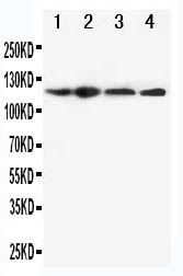
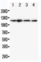
- Western blot analysis of -CIAS1/NALP3 using anti- -CIAS1/NALP3 antibody (PA1665). Electrophoresis was performed on a 5-20% SDS-PAGE gel at 70V (Stacking gel) / 90V (Resolving gel) for 2-3 hours. The sample well of each lane was loaded with 50ug of sample under reducing conditions. Lane 1: HEP-2 Cell Lysate Lane 2: A549 Cell Lysate Lane 3: U87 Cell Lysate Lane 4: CEM Cell Lysate After Electrophoresis, proteins were transferred to a Nitrocellulose membrane at 150mA for 50-90 minutes. Blocked the membrane with 5% Non-fat Milk/ TBS for 1.5 hour at RT. The membrane was incubated with rabbit anti- -CIAS1/NALP3 antigen affinity purified polyclonal antibody (Catalog # PA1665) at 0.5 μg/mL overnight at 4°C, then washed with TBS-0.1%Tween 3 times with 5 minutes each and probed with a goat anti-rabbit IgG-HRP secondary antibody at a dilution of 1:10000 for 1.5 hour at RT. The signal is developed using an Enhanced Chemiluminescent detection (ECL) kit (Catalog # EK1002) with Tanon 5200 system. A specific band was detected for -CIAS1/NALP3 at approximately 118KD. The expected band size for -CIAS1/NALP3 is at 118KD.
- Additional image