Explore
 Validate
 Learn
Learn Western blot
 Immunohistochemistry
Antibody data
- Antibody Data
- Antigen structure
- References [3]
- Comments [0]
- Validations
- Immunohistochemistry [1]
Submit
Validation data
Reference
Comment
Report error
- Product number
- HPA016604 - Provider product page

- Provider
- Atlas Antibodies
- Proper citation
- Atlas Antibodies Cat#HPA016604, RRID:AB_1857859
- Product name
- Anti-TCL1A
- Antibody type
- Polyclonal
- Description
- Polyclonal Antibody against Human TCL1A, Gene description: T-cell leukemia/lymphoma 1A, Alternative Gene Names: TCL1, Validated applications: IHC, WB, Uniprot ID: P56279, Storage: Store at +4°C for short term storage. Long time storage is recommended at -20°C.
- Reactivity
- Human
- Host
- Rabbit
- Conjugate
- Unconjugated
- Isotype
- IgG
- Vial size
- 100 µl
- Concentration
- 0.2 mg/ml
- Storage
- Store at +4°C for short term storage. Long time storage is recommended at -20°C.
- Handling
- The antibody solution should be gently mixed before use.
Submitted references Exosomal mRNA Cargo are biomarkers of tumor and immune cell populations in pediatric osteosarcoma
The Transcriptomic and Proteomic Landscapes of Bone Marrow and Secondary Lymphoid Tissues
IGHV3‐21 gene usage is associated with high TCL1 expression in chronic lymphocytic leukemia
Ong J, Jalaludin N, Wong M, Tan S, Angelina C, Sukhatme S, Yeo T, Lim C, Lee Y, Soh S, Lim T, Tay T, Chang K, Chen Z, Loh A
Translational Oncology 2024;46
Translational Oncology 2024;46
The Transcriptomic and Proteomic Landscapes of Bone Marrow and Secondary Lymphoid Tissues
Rameshwar P, Andersson S, Nilsson K, Fagerberg L, Hallström B, Sundström C, Danielsson A, Edlund K, Uhlen M, Asplund A
PLoS ONE 2014;9(12):e115911
PLoS ONE 2014;9(12):e115911
IGHV3‐21 gene usage is associated with high TCL1 expression in chronic lymphocytic leukemia
Mansouri M, Sevov M, Åleskog A, Jondal M, Merup M, Sundström C, Osorio L, Rosenquist R
European Journal of Haematology 2010;84(2):109-116
European Journal of Haematology 2010;84(2):109-116
No comments: Submit comment
Supportive validation
- Submitted by
- Atlas Antibodies (provider)
- Enhanced method
- Orthogonal validation
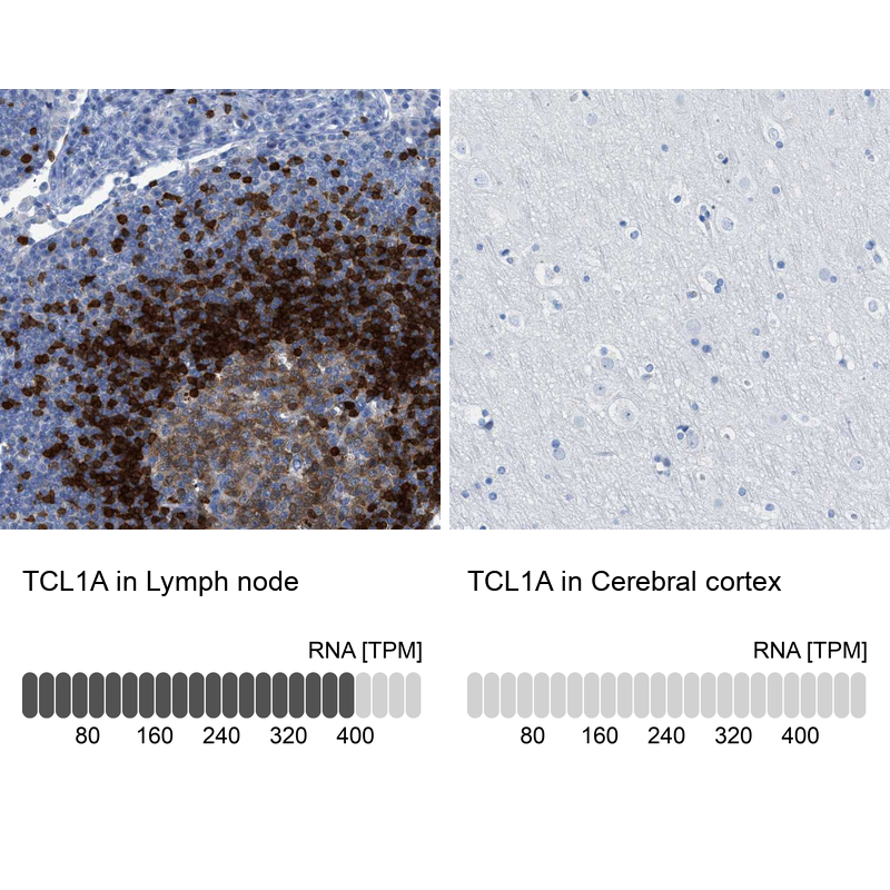
- Main image
- Experimental details
- Immunohistochemistry analysis in human lymph node and cerebral cortex tissues using HPA016604 antibody. Corresponding TCL1A RNA-seq data are presented for the same tissues.
- Sample type
- Human
- Protocol
- Protocol