Explore
 Validate
 Learn
Learn Western blot
Antibody data
- Antibody Data
- Antigen structure
- References [1]
- Comments [0]
- Validations
- Western blot [1]
- Immunohistochemistry [2]
- Flow cytometry [2]
Submit
Validation data
Reference
Comment
Report error
- Product number
- PA5-35315 - Provider product page

- Provider
- Invitrogen Antibodies
- Product name
- Transthyretin Polyclonal Antibody
- Antibody type
- Polyclonal
- Antigen
- Synthetic peptide
- Description
- This antibody is predicted to react with non-human primate based on sequence homology.
- Reactivity
- Human, Rat
- Host
- Rabbit
- Isotype
- IgG
- Vial size
- 400 μL
- Concentration
- 0.5 mg/mL
- Storage
- Store at 4°C short term. For long term storage, store at -20°C, avoiding freeze/thaw cycles.
Submitted references Transthyretin amyloid fibrils alter primary fibroblast structure, function, and inflammatory gene expression.
Dittloff KT, Iezzi A, Zhong JX, Mohindra P, Desai TA, Russell B
American journal of physiology. Heart and circulatory physiology 2021 Jul 1;321(1):H149-H160
American journal of physiology. Heart and circulatory physiology 2021 Jul 1;321(1):H149-H160
No comments: Submit comment
Supportive validation
- Submitted by
- Invitrogen Antibodies (provider)
- Main image
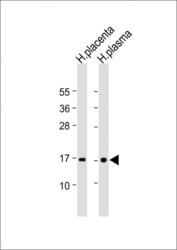
- Experimental details
- Western blot analysis of Transthyretin in various lysates. Samples were incubated with Transthyretin polyclonal antibody (Product # PA5-35315) followed by Goat Anti-Rabbit IgG, (H+L), Peroxidase conjugated at a dilution of 1:10,000. Lysates/proteins: 20 µg per lane. Lane 1: Human placenta lysate; Lane 2: Human plasma lysate. Predicted band size: 16 kDa. Blocking/Dilution buffer: 5% NFDM/TBST.

Supportive validation
- Submitted by
- Invitrogen Antibodies (provider)
- Main image
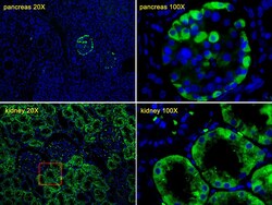
- Experimental details
- Immunohistochemistry analysis of Transthyretin in Human pancreas tissues and Human kidney tissues. Samples were incubated with Transthyretin polyclonal antibody (Product # PA5-35315) using a dilution of 0.0590277777777778 followed by Alexa Fluor 488-conjugated goat anti-rabbit lgG at a dilution of 1:400 (green). DAPI was used to stain the cell nuclear (blue).

- Submitted by
- Invitrogen Antibodies (provider)
- Main image
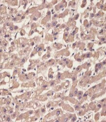
- Experimental details
- Immunohistochemistry analysis of Transthyretin in paraformaldehyde-fixed, paraffin-embedded human liver tissue sections. Samples were incubated with Transthyretin polyclonal antibody (Product # PA5-35315) using a dilution of 1:25 for 1 hours at 37°C followed by an undiluted biotinylated goat polyvalent antibody. Tissue was fixed with formaldehyde and blocked with 3% BSA for 0.5 hour at room temperature; antigen retrieval was by heat mediation with a citrate buffer (pH 6).

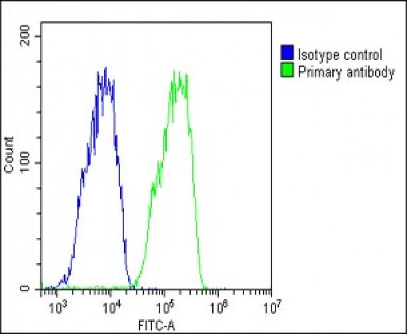
Supportive validation
- Submitted by
- Invitrogen Antibodies (provider)
- Main image
- Experimental details
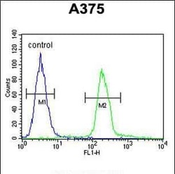
- Flow cytometry analysis of TTR in A375 cells (right) compared to a negative control (left) using a TTR polyclonal antibody (Product # PA5-35315) followed by detection using a FITC-conjugated goat-anti-rabbit secondary antibody.

- Submitted by
- Invitrogen Antibodies (provider)
- Main image

- Experimental details
- Flow cytometry of (overlay histogram) of Transthyretin in HepG2 cells (green line). Samples were incubated with Transthyretin polyclonal antibody (Product # PA5-35315) using a dilution of 1:25 dilution for 60 min at 37°C followed by Goat-Anti-Rabbit IgG, DyLight® 488 Conjugated Highly Cross-Adsorbed at 1:200 dilution for 40 min at 37°C. The cells were fixed with 2% paraformaldehyde (10 min) and then permeabilized with 90% methanol for 10 min. The cells were then incubated in 2% bovine serum albumin to block non-specific protein-protein interactions followed by the primary antibody. Isotype control antibody (blue line) was rabbit IgG1 (1 μg/1x10^6 cells) used under the same conditions. Acquisition of >10, 000 events was performed.