Explore
 Validate
 Learn
Learn Western blot
 Immunocytochemistry
Antibody data
- Antibody Data
- Antigen structure
- References [1]
- Comments [0]
- Validations
- Immunocytochemistry [4]
- Immunohistochemistry [5]
- Flow cytometry [2]
- Other assay [1]
Submit
Validation data
Reference
Comment
Report error
- Product number
- MA5-32634 - Provider product page

- Provider
- Invitrogen Antibodies
- Product name
- Transthyretin Recombinant Rabbit Monoclonal Antibody (JM11-43)
- Antibody type
- Monoclonal
- Antigen
- Synthetic peptide
- Description
- Recombinant rabbit monoclonal antibodies are produced using in vitro expression systems. The expression systems are developed by cloning in the specific antibody DNA sequences from immunoreactive rabbits. Then, individual clones are screened to select the best candidates for production. The advantages of using recombinant rabbit monoclonal antibodies include: better specificity and sensitivity, lot-to-lot consistency, animal origin-free formulations, and broader immunoreactivity to diverse targets due to larger rabbit immune repertoire.
- Reactivity
- Human
- Host
- Rabbit
- Isotype
- IgG
- Antibody clone number
- JM11-43
- Vial size
- 100 μL
- Concentration
- 1 mg/mL
- Storage
- Store at 4°C short term. For long term storage, store at -20°C, avoiding freeze/thaw cycles.
Submitted references Integrative Multi-Omics Analysis in Calcific Aortic Valve Disease Reveals a Link to the Formation of Amyloid-Like Deposits.
Heuschkel MA, Skenteris NT, Hutcheson JD, van der Valk DD, Bremer J, Goody P, Hjortnaes J, Jansen F, Bouten CVC, van den Bogaerdt A, Matic L, Marx N, Goettsch C
Cells 2020 Sep 24;9(10)
Cells 2020 Sep 24;9(10)
No comments: Submit comment
Supportive validation
- Submitted by
- Invitrogen Antibodies (provider)
- Main image

- Experimental details
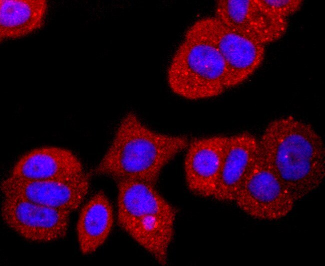
- Immunocytochemical analysis of Transthyretin in A549 cells using a Transthyretin Monoclonal antibody (Product # MA5-32634) as seen in red. The nuclear counter stain is DAPI (blue). Cells were fixed in paraformaldehyde, permeabilised with 0.25% Triton X100/PBS.

- Submitted by
- Invitrogen Antibodies (provider)
- Main image

- Experimental details
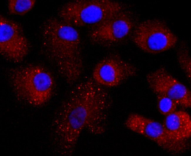
- Immunocytochemical analysis of Transthyretin in MCF-7 cells using a Transthyretin Monoclonal antibody (Product # MA5-32634) as seen in red. The nuclear counter stain is DAPI (blue). Cells were fixed in paraformaldehyde, permeabilised with 0.25% Triton X100/PBS.
- Submitted by
- Invitrogen Antibodies (provider)
- Main image

- Experimental details
- Immunocytochemical analysis of Transthyretin in A549 cells using a Transthyretin Monoclonal antibody (Product # MA5-32634) as seen in red. The nuclear counter stain is DAPI (blue). Cells were fixed in paraformaldehyde, permeabilised with 0.25% Triton X100/PBS.
- Submitted by
- Invitrogen Antibodies (provider)
- Main image

- Experimental details
- Immunocytochemical analysis of Transthyretin in MCF-7 cells using a Transthyretin Monoclonal antibody (Product # MA5-32634) as seen in red. The nuclear counter stain is DAPI (blue). Cells were fixed in paraformaldehyde, permeabilised with 0.25% Triton X100/PBS.

Supportive validation
- Submitted by
- Invitrogen Antibodies (provider)
- Main image

- Experimental details
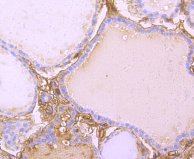
- Immunohistochemical analysis of Transthyretin of paraffin-embedded Human thyroid tissue using a Transthyretin Monoclonal antibody (Product #MA5-32634). Counter stained with hematoxylin.
- Submitted by
- Invitrogen Antibodies (provider)
- Main image
- Experimental details
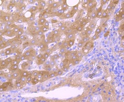
- Immunohistochemical analysis of Transthyretin of paraffin-embedded Human liver cancer tissue using a Transthyretin Monoclonal antibody (Product #MA5-32634). Counter stained with hematoxylin.

- Submitted by
- Invitrogen Antibodies (provider)
- Main image
- Experimental details
- Immunohistochemical analysis of Transthyretin of paraffin-embedded Human liver tissue using a Transthyretin Monoclonal antibody (Product #MA5-32634). Counter stained with hematoxylin.
- Submitted by
- Invitrogen Antibodies (provider)
- Main image

- Experimental details
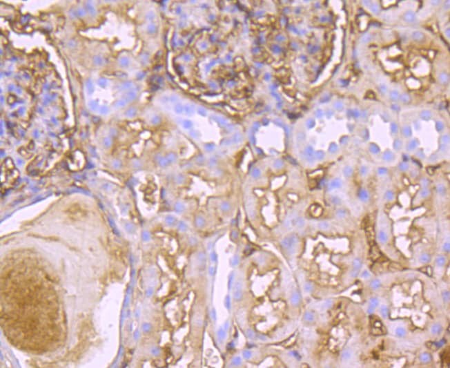
- Immunohistochemical analysis of Transthyretin of paraffin-embedded Human kidney tissue using a Transthyretin Monoclonal antibody (Product #MA5-32634). Counter stained with hematoxylin.
- Submitted by
- Invitrogen Antibodies (provider)
- Main image

- Experimental details
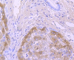
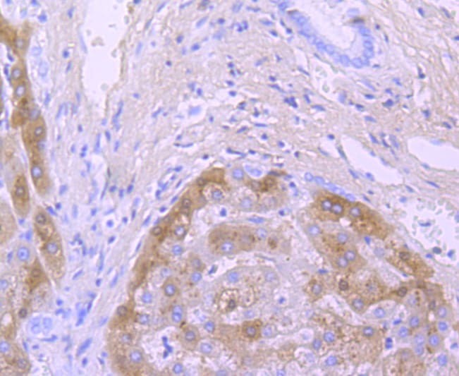
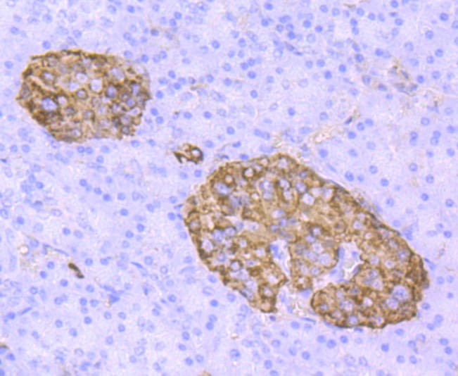
- Immunohistochemical analysis of Transthyretin of paraffin-embedded Human pancreas tissue using a Transthyretin Monoclonal antibody (Product #MA5-32634). Counter stained with hematoxylin.
Supportive validation
- Submitted by
- Invitrogen Antibodies (provider)
- Main image
- Experimental details
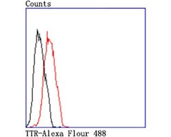
- Flow Cytometric analysis of Transthyretin in MCF-7 cells using a Transthyretin Monoclonal Antibody (Product # MA5-32634) at a dilution of 1:50, as seen in red compared with an unlabelled control (cells without incubation with primary antibody; black). Alexa Fluor 488-conjugated goat anti rabbit IgG was used as the secondary antibody.

- Submitted by
- Invitrogen Antibodies (provider)
- Main image

- Experimental details
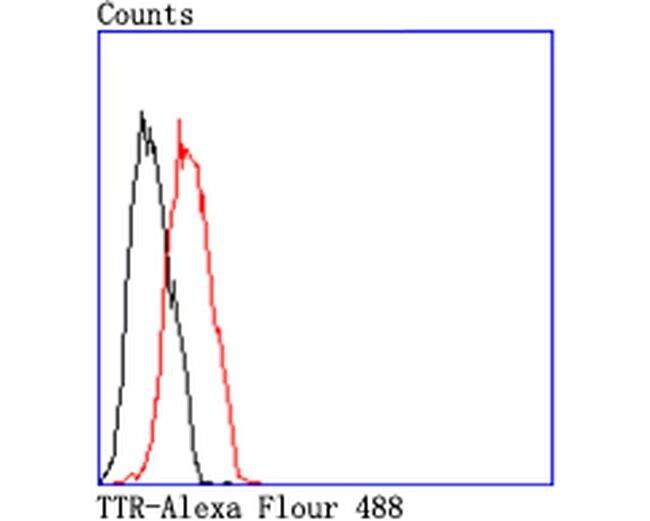
- Flow Cytometric analysis of Transthyretin in MCF-7 cells using a Transthyretin Monoclonal Antibody (Product # MA5-32634) at a dilution of 1:50, as seen in red compared with an unlabelled control (cells without incubation with primary antibody; black). Alexa Fluor 488-conjugated goat anti rabbit IgG was used as the secondary antibody.
Supportive validation
- Submitted by
- Invitrogen Antibodies (provider)
- Main image
- Experimental details
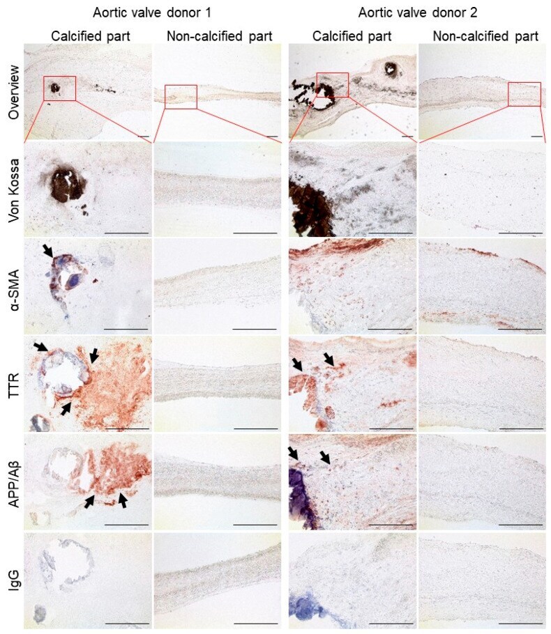
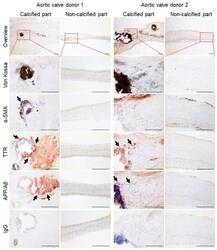
- Figure 4 Human calcified aortic valves are transthyretin (TTR) and amyloid precursor protein (APP)/beta-amyloid (Abeta)-immunoreactive. Aortic valves from patients with aortic stenosis were divided into the calcified and non-calcified portions. Calcification was visualized by von Kossa staining. Immunohistochemistry was performed for smooth muscle alpha-actin (alphaSMA), TTR, and APP/Abeta. Immunoglobulin G (IgG) served as control. One leaflet from each of two donors is shown. The first row shows an overview of the tissue and indicates the respective area of the magnified images. The arrows indicate positive signal. Bar: 200 mum.