 Explore
 Validate
 Learn
Learn Western blot
Antibody data
- Antibody Data
- Antigen structure
- References [3]
- Comments [0]
- Validations
- Western blot [6]
- Other assay [3]
Submit
Validation data
Reference
Comment
Report error
- Product number
- PA5-27220 - Provider product page

- Provider
- Invitrogen Antibodies
- Product name
- Transthyretin Polyclonal Antibody
- Antibody type
- Polyclonal
- Antigen
- Recombinant full-length protein
- Description
- Recommended positive controls: Human plasma, Mouse liver, rat plasma. Predicted reactivity: Mouse (81%), Rat (82%), Cat (85%), Pig (87%), Sheep (85%), Rhesus Monkey (93%), Chimpanzee (97%), Bovine (85%). Store product as a concentrated solution. Centrifuge briefly prior to opening the vial.
- Reactivity
- Human, Mouse, Rat
- Host
- Rabbit
- Isotype
- IgG
- Vial size
- 100 µL
- Concentration
- 1.58 mg/mL
- Storage
- Store at 4°C short term. For long term storage, store at -20°C, avoiding freeze/thaw cycles.
Submitted references Proteomic Exploration of Plasma Exosomes and Other Small Extracellular Vesicles in Pediatric Hodgkin Lymphoma: A Potential Source of Biomarkers for Relapse Occurrence.
Transthyretin expression in the postischemic brain.
Phaseic Acid, an Endogenous and Reversible Inhibitor of Glutamate Receptors in Mouse Brain.
Repetto O, Lovisa F, Elia C, Enderle D, Romanato F, Buffardi S, Sala A, Pillon M, Steffan A, Burnelli R, Mussolin L, Mascarin M, De Re V
Diagnostics (Basel, Switzerland) 2021 May 21;11(6)
Diagnostics (Basel, Switzerland) 2021 May 21;11(6)
Transthyretin expression in the postischemic brain.
Talhada D, Gonçalves I, Gomes JC, Saraiva MJ, Reis Santos C, Ruscher K
PloS one 2019;14(9):e0221555
PloS one 2019;14(9):e0221555
Phaseic Acid, an Endogenous and Reversible Inhibitor of Glutamate Receptors in Mouse Brain.
Hou ST, Jiang SX, Zaharia LI, Han X, Benson CL, Slinn J, Abrams SR
The Journal of biological chemistry 2016 Dec 30;291(53):27007-27022
The Journal of biological chemistry 2016 Dec 30;291(53):27007-27022
No comments: Submit comment
Supportive validation
- Submitted by
- Invitrogen Antibodies (provider)
- Main image

- Experimental details
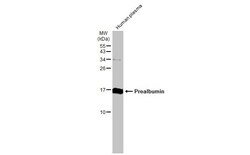
- Western blot analysis of Transthyretin in Human tissue extract (30 µg). Samples was separated by 15% SDS-PAGE and the membrane was probed with Transthyretin Polyclonal antibody (Product # PA5-27220) at a dilution of 1:500.
- Submitted by
- Invitrogen Antibodies (provider)
- Main image

- Experimental details
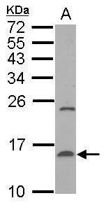
- Western blot analysis of Transthyretin using 50 µg of mouse liver lysate. Samples were loaded onto a 15% SDS-PAGE gel and probed with a Transthyretin polyclonal antibody (Product # PA5-27220) at a dilution of 1:500.
- Submitted by
- Invitrogen Antibodies (provider)
- Main image
- Experimental details
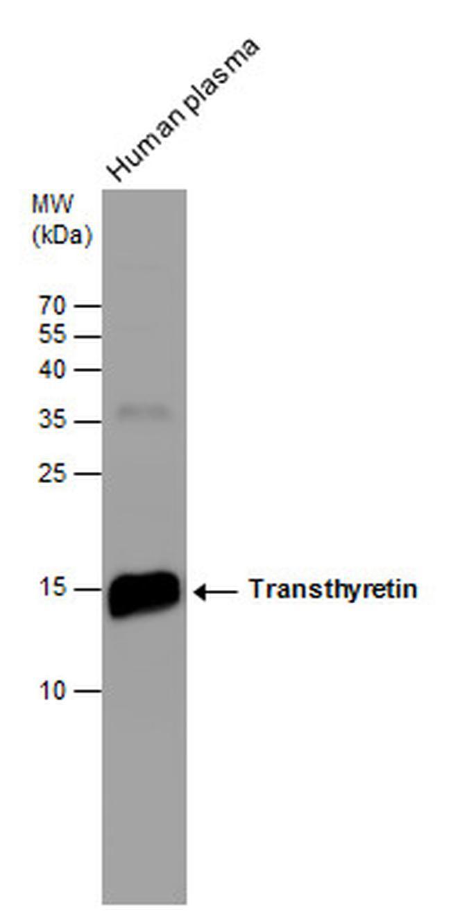
- Western blot analysis of Transthyretin was performed by separating 30 µg of human plasma by 15% SDS-PAGE. Proteins were transferred to a membrane and probed with a Transthyretin Polyclonal Antibody (Product # PA5-27220) at a dilution of 1:500. The HRP-conjugated anti-rabbit IgG antibody was used to detect the primary antibody.

- Submitted by
- Invitrogen Antibodies (provider)
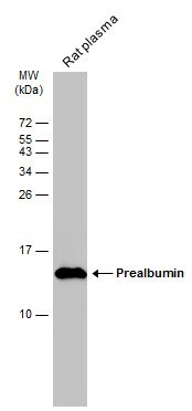
- Main image
- Experimental details
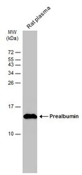
- Western blot analysis of Transthyretin was performed by separating 50 µg of rat plasma by 15% SDS-PAGE. Proteins were transferred to a membrane and probed with a Transthyretin Polyclonal Antibody (Product # PA5-27220) at a dilution of 1:500. The HRP-conjugated anti-rabbit IgG antibody was used to detect the primary antibody.
- Submitted by
- Invitrogen Antibodies (provider)
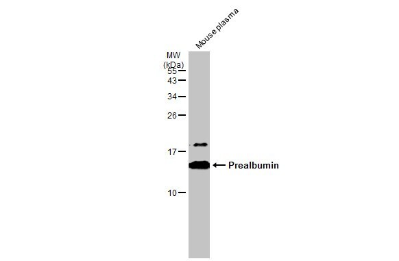
- Main image
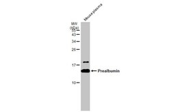
- Experimental details
- Western Blot using Transthyretin Polyclonal Antibody (Product # PA5-27220). Mouse plasma (50 µg) was separated by 15% SDS-PAGE, and the membrane was blotted with Transthyretin Polyclonal Antibody (Product # PA5-27220) diluted at 1:500. The HRP-conjugated anti-rabbit IgG antibody was used to detect the primary antibody.
- Submitted by
- Invitrogen Antibodies (provider)
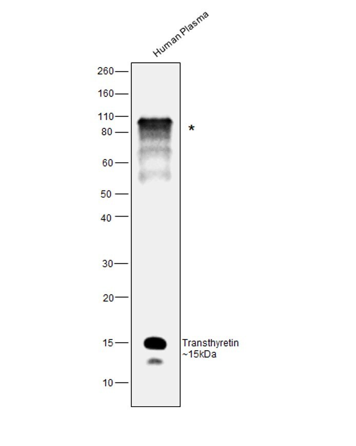
- Main image

- Experimental details
- Western blot was performed using Anti-Transthyretin Polyclonal Antibody (Product # PA5-27220) and a 15 kDa band along with lower band as reported isoform and an uncharacterized band (*) at ~80 kDa corresponding to Transthyretin was observed in plasma sample, 2 µL lysate of Human Plasma (Lane 1) were electrophoresed using NuPAGE™ 4-12% Bis-Tris Protein Gel (Product # NP0321BOX). Resolved proteins were then transferred onto a Nitrocellulose membrane (Product # IB23002) by iBlot® 2 Dry Blotting System (Product # IB21001). The blot was probed with the primary antibody (1:1000 dilution) and detected by chemiluminescence with Goat anti-Rabbit IgG (H+L) Superclonal™ Recombinant Secondary Antibody, HRP (Product # A27036, 1:4000 dilution) using the iBright FL 1000 (Product # A32752). Chemiluminescent detection was performed using Novex® ECL Chemiluminescent Substrate Reagent Kit (Product # WP20005).
Supportive validation
- Submitted by
- Invitrogen Antibodies (provider)
- Main image
- Experimental details
- NULL
- Submitted by
- Invitrogen Antibodies (provider)
- Main image
- Experimental details
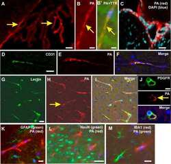
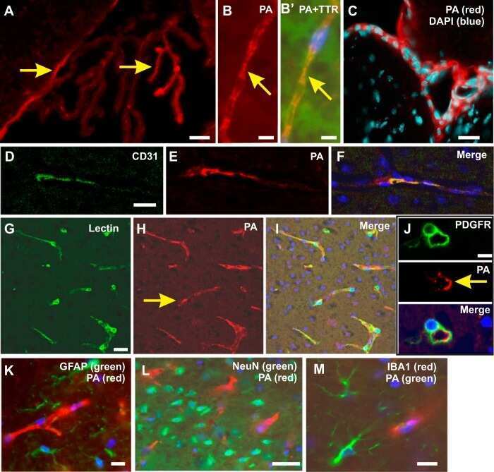
- Fig 2 Immunohistochemistry in the postischemic brain. Scattered cells were immunoreactive for TTR in the IC and PI 14 days after ischemic stroke. Low magnification overview of the ischemic territory using 1:1000 anti-TTR rabbit antibody (ThermoScientific cat# PA5-27220, Rockford, USA) (A) without and (D) with BP. (B, E and E') Higher magnification micrographs of images (A) showing cells immunoreactivity for TTR (white arrows). (C) Epithelial cells from CP were positive for TTR and (F) partially blocked with BP. (G) Staining for TTR (green, AF488) and with higher magnification in (J) together with DAPI (H). (J) Epithelial cells from CP from the same section. Scale bars: A , C , D and F-- 100 mum, B and E-- 20 mum and E' - 10 mum. BP-Blocking peptide; CP--Choroid plexus; IC--Infarct core; PI-Peri-infarct area; TTR-Transthyretin; LV-Lateral ventricle.

- Submitted by
- Invitrogen Antibodies (provider)
- Main image
- Experimental details
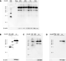
- Fig 5 Evaluation of a chicken TTR antibody and TTR levels in brain parenchyma. (A) In contrast to protein extracts obtained from ischemic territory (infarct core-peri-infarct area) at 24 hours, 48 hours, 7 days and 14 days after PT, the monomeric form of TTR is found in samples of mouse serum, used as positive control. (B) TTR in human and mouse serum and absence of the protein in the cortical ischemic territory 14 days after PT. Instead, a band of approximately 60 to 65 kDa is found, not corresponding to the TTR protein (C) Blocking of specific TTR bands by preincubation with a specific TTR peptide. (D) Western blot performed without primary antibody incubation. HS-Human serum; M-Marker; MS-mouse serum; PT-Photothrombosis; TTR-Transthyretin; cx-cortex.
