 Explore
 Validate
 Learn
Learn Flow cytometry
Antibody data
- Antibody Data
- Antigen structure
- References [4]
- Comments [0]
- Validations
- Flow cytometry [1]
Submit
Validation data
Reference
Comment
Report error
- Product number
- FAB5204P-025 - Provider product page

- Provider
- R&D Systems
- Product name
- Human CX3CR1 PE-conjugated Antibody
- Antibody type
- Monoclonal
- Description
- Protein A or G purified from hybridoma culture supernatant. Detects human CX3CR1 in direct ELISAs.
- Reactivity
- Human
- Host
- Mouse
- Antigen sequence
NP_001328- Isotype
- IgG
- Antibody clone number
- 528728
- Vial size
- 25 Tests
- Storage
- Protect from light. Do not freeze. 12 months from date of receipt, 2 to 8 °C as supplied.
Submitted references Phenotypic changes of peripheral blood mononuclear cells upon corticosteroid treatment in idiopathic intermediate uveitis.
CX3CL1/CX3CR1 and CCL2/CCR2 chemokine/chemokine receptor complex in patients with AMD.
CD56(bright)perforin(low) noncytotoxic human NK cells are abundant in both healthy and neoplastic solid tissues and recirculate to secondary lymphoid organs via afferent lymph.
Changes of peripheral TGF-β1 depend on monocytes-derived macrophages in Huntington disease.
Walscheid K, Weinhage T, Foell D, Heinz C, Kasper M, Heiligenhaus A
Clinical immunology (Orlando, Fla.) 2016 Oct 28;
Clinical immunology (Orlando, Fla.) 2016 Oct 28;
CX3CL1/CX3CR1 and CCL2/CCR2 chemokine/chemokine receptor complex in patients with AMD.
Falk MK, Singh A, Faber C, Nissen MH, Hviid T, Sørensen TL
PloS one 2014;9(12):e112473
PloS one 2014;9(12):e112473
CD56(bright)perforin(low) noncytotoxic human NK cells are abundant in both healthy and neoplastic solid tissues and recirculate to secondary lymphoid organs via afferent lymph.
Carrega P, Bonaccorsi I, Di Carlo E, Morandi B, Paul P, Rizzello V, Cipollone G, Navarra G, Mingari MC, Moretta L, Ferlazzo G
Journal of immunology (Baltimore, Md. : 1950) 2014 Apr 15;192(8):3805-15
Journal of immunology (Baltimore, Md. : 1950) 2014 Apr 15;192(8):3805-15
Changes of peripheral TGF-β1 depend on monocytes-derived macrophages in Huntington disease.
Di Pardo A, Alberti S, Maglione V, Amico E, Cortes EP, Elifani F, Battaglia G, Busceti CL, Nicoletti F, Vonsattel JP, Squitieri F
Molecular brain 2013 Dec 13;6:55
Molecular brain 2013 Dec 13;6:55
No comments: Submit comment
Supportive validation
- Submitted by
- R&D Systems (provider)
- Main image
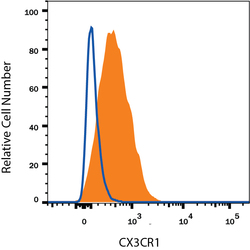
- Experimental details
- Detection of CX3CR1 in Human Blood Monocytes by Flow Cytometry. Human peripheral blood monocytes were stained with Mouse Anti-Human CX3CR1 PE-conjugated Monoclonal Antibody (Catalog # FAB5204P, filled histogram) or isotype control antibody (Catalog # IC002P, open histogram). View our protocol for Staining Membrane-associated Proteins.
