 Explore
 Validate
 Learn
Learn Flow cytometry
Antibody data
- Antibody Data
- Antigen structure
- References [5]
- Comments [0]
- Validations
- Flow cytometry [1]
Submit
Validation data
Reference
Comment
Report error
- Product number
- 13-6099-80 - Provider product page

- Provider
- Invitrogen Antibodies
- Product name
- Anti-CX3CR1 Monoclonal Antibody (2A9-1), Biotin, eBioscience™
- Antibody type
- Monoclonal
- Antigen
- Other
- Description
- Description: This 2A9-1 monoclonal antibody reacts with human CX3CR1, which is the receptor for fractalkine, a transmembrane chemokine of the CX3C family. CX3CR1 is a seven transmembrane G protein-coupled receptor expressed on CD16+ NK cells, T cells (e.g. CD8+, CD4+, and gamma/delta), and monocytes. In non-immune cells, CX3CR1 has been found on osteoclast precursors and microglia. Little to no CX3CR1 surface expression can be detected on B cells and granulocytes. Together, fractalkine and its receptor mediate cell-cell adhesion and chemotaxis of NK cells, T cells, and monocytes. The expression of CX3CR1 has also been correlated with high levels of intracellular perforin and granzyme B. Applications Reported: This 2A9-1 antibody has been reported for use in flow cytometric analysis. Applications Tested: This 2A9-1 antibody has been tested by flow cytometric analysis of normal human peripheral blood cells. This can be used at less than or equal to 0.25 µg per test. A test is defined as the amount (µg) of antibody that will stain a cell sample in a final volume of 100 µL. Cell number should be determined empirically but can range from 10^5 to 10^8 cells/test. It is recommended that the antibody be carefully titrated for optimal performance in the assay of interest. Filtration: 0.2 µm post-manufacturing filtered.
- Reactivity
- Human
- Host
- Rat
- Conjugate
- Biotin
- Isotype
- IgG
- Antibody clone number
- 2A9-1
- Vial size
- 25 µg
- Concentration
- 0.5 mg/mL
- Storage
- 4° C, store in dark, DO NOT FREEZE!
Submitted references Microglia innately develop within cerebral organoids.
Modulation of Hematopoietic Lineage Specification Impacts TREM2 Expression in Microglia-Like Cells Derived From Human Stem Cells.
Pivotal Role for CD16+ Monocytes in Immune Surveillance of the Central Nervous System.
The CD14+CD16+ inflammatory monocyte subset displays increased mitochondrial activity and effector function during acute Plasmodium vivax malaria.
Dual functions of fractalkine/CX3C ligand 1 in trafficking of perforin+/granzyme B+ cytotoxic effector lymphocytes that are defined by CX3CR1 expression.
Ormel PR, Vieira de Sá R, van Bodegraven EJ, Karst H, Harschnitz O, Sneeboer MAM, Johansen LE, van Dijk RE, Scheefhals N, Berdenis van Berlekom A, Ribes Martínez E, Kling S, MacGillavry HD, van den Berg LH, Kahn RS, Hol EM, de Witte LD, Pasterkamp RJ
Nature communications 2018 Oct 9;9(1):4167
Nature communications 2018 Oct 9;9(1):4167
Modulation of Hematopoietic Lineage Specification Impacts TREM2 Expression in Microglia-Like Cells Derived From Human Stem Cells.
Amos PJ, Fung S, Case A, Kifelew J, Osnis L, Smith CL, Green K, Naydenov A, Aloi M, Hubbard JJ, Ramakrishnan A, Garden GA, Jayadev S
ASN neuro 2017 Jul-Aug;9(4):1759091417716610
ASN neuro 2017 Jul-Aug;9(4):1759091417716610
Pivotal Role for CD16+ Monocytes in Immune Surveillance of the Central Nervous System.
Waschbisch A, Schröder S, Schraudner D, Sammet L, Weksler B, Melms A, Pfeifenbring S, Stadelmann C, Schwab S, Linker RA
Journal of immunology (Baltimore, Md. : 1950) 2016 Feb 15;196(4):1558-67
Journal of immunology (Baltimore, Md. : 1950) 2016 Feb 15;196(4):1558-67
The CD14+CD16+ inflammatory monocyte subset displays increased mitochondrial activity and effector function during acute Plasmodium vivax malaria.
Antonelli LR, Leoratti FM, Costa PA, Rocha BC, Diniz SQ, Tada MS, Pereira DB, Teixeira-Carvalho A, Golenbock DT, Gonçalves R, Gazzinelli RT
PLoS pathogens 2014 Sep;10(9):e1004393
PLoS pathogens 2014 Sep;10(9):e1004393
Dual functions of fractalkine/CX3C ligand 1 in trafficking of perforin+/granzyme B+ cytotoxic effector lymphocytes that are defined by CX3CR1 expression.
Nishimura M, Umehara H, Nakayama T, Yoneda O, Hieshima K, Kakizaki M, Dohmae N, Yoshie O, Imai T
Journal of immunology (Baltimore, Md. : 1950) 2002 Jun 15;168(12):6173-80
Journal of immunology (Baltimore, Md. : 1950) 2002 Jun 15;168(12):6173-80
No comments: Submit comment
Supportive validation
- Submitted by
- Invitrogen Antibodies (provider)
- Main image
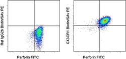
- Experimental details
- Staining of normal human peripheral blood cells with 0.125 µg of Rat IgG2b Isotype Control Biotin (Product # 13-4031-82) (left) or 0.125 µg of Anti-Human CX3CR1 Biotin (right) followed by Streptavidin PE (Product # 12-4317-87). Samples were subsequently stained intracellularly with Anti-Human Perforin FITC (Product # 11-9994-42). Cells were gated on CD16+CD56+ using Anti-Human CD16 eFluor® 450 (Product # 48-0168-42) and Anti-Human CD56 APC.
- Conjugate
- Biotin
