Explore
 Validate
 Learn
Learn Flow cytometry
Antibody data
- Antibody Data
- Antigen structure
- References [4]
- Comments [0]
- Validations
- Flow cytometry [1]
- Other assay [3]
Submit
Validation data
Reference
Comment
Report error
- Product number
- 25-6099-42 - Provider product page

- Provider
- Invitrogen Antibodies
- Product name
- CX3CR1 Monoclonal Antibody (2A9-1), PE-Cyanine7, eBioscience™
- Antibody type
- Monoclonal
- Antigen
- Other
- Description
- Description: This 2A9-1 monoclonal antibody reacts with human CX3CR1, which is the receptor for fractalkine, a transmembrane chemokine of the CX3C family. CX3CR1 is a seven transmembrane G protein-coupled receptor expressed on CD16+ NK cells, T cells (e.g. CD8+, CD4+, and gamma/delta), and monocytes. In non-immune cells, CX3CR1 has been found on osteoclast precursors and microglia. Little to no CX3CR1 surface expression can be detected on B cells and granulocytes. Together, fractalkine and its receptor mediate cell-cell adhesion and chemotaxis of NK cells, T cells, and monocytes. The expression of CX3CR1 has also been correlated with high levels of intracellular perforin and granzyme B. Applications Reported: This 2A9-1 antibody has been reported for use in flow cytometric analysis. Applications Tested: This 2A9-1 antibody has been pre-titrated and tested by flow cytometric analysis of normal human peripheral blood cells. This can be used at 5 µL (0.125 µg) per test. A test is defined as the amount (µg) of antibody that will stain a cell sample in a final volume of 100 µL. Cell number should be determined empirically but can range from 10^5 to 10^8 cells/test. Light sensitivity: This tandem dye is sensitive to photo-induced oxidation. Please protect this vial and stained samples from light. Fixation: Samples can be stored in IC Fixation Buffer (Product # 00-822-49) (100 µL of cell sample + 100 µL of IC Fixation Buffer) or 1-step Fix/Lyse Solution (Product # 00-5333-54) for up to 3 days in the dark at 4°C with minimal impact on brightness and FRET efficiency/compensation. Some generalizations regarding fluorophore performance after fixation can be made, but clone specific performance should be determined empirically. Excitation: 488-561 nm; Emission: 775 nm; Laser: Blue Laser, Green Laser, Yellow-Green Laser. Filtration: 0.2 µm post-manufacturing filtered.
- Reactivity
- Human
- Host
- Rat
- Isotype
- IgG
- Antibody clone number
- 2A9-1
- Vial size
- 100 Tests
- Concentration
- 5 µL/Test
- Storage
- 4° C, store in dark, DO NOT FREEZE!
Submitted references Distinct gene expression patterns for CD14++ and CD16++ monocytes in preeclampsia.
Microglia innately develop within cerebral organoids.
Modulation of Hematopoietic Lineage Specification Impacts TREM2 Expression in Microglia-Like Cells Derived From Human Stem Cells.
Dual functions of fractalkine/CX3C ligand 1 in trafficking of perforin+/granzyme B+ cytotoxic effector lymphocytes that are defined by CX3CR1 expression.
Vishnyakova P, Kuznetsova M, Poltavets A, Fomina M, Kiseleva V, Muminova K, Potapova A, Khodzhaeva Z, Pyregov A, Trofimov D, Elchaninov A, Sukhikh G, Fatkhudinov T
Scientific reports 2022 Sep 14;12(1):15469
Scientific reports 2022 Sep 14;12(1):15469
Microglia innately develop within cerebral organoids.
Ormel PR, Vieira de Sá R, van Bodegraven EJ, Karst H, Harschnitz O, Sneeboer MAM, Johansen LE, van Dijk RE, Scheefhals N, Berdenis van Berlekom A, Ribes Martínez E, Kling S, MacGillavry HD, van den Berg LH, Kahn RS, Hol EM, de Witte LD, Pasterkamp RJ
Nature communications 2018 Oct 9;9(1):4167
Nature communications 2018 Oct 9;9(1):4167
Modulation of Hematopoietic Lineage Specification Impacts TREM2 Expression in Microglia-Like Cells Derived From Human Stem Cells.
Amos PJ, Fung S, Case A, Kifelew J, Osnis L, Smith CL, Green K, Naydenov A, Aloi M, Hubbard JJ, Ramakrishnan A, Garden GA, Jayadev S
ASN neuro 2017 Jul-Aug;9(4):1759091417716610
ASN neuro 2017 Jul-Aug;9(4):1759091417716610
Dual functions of fractalkine/CX3C ligand 1 in trafficking of perforin+/granzyme B+ cytotoxic effector lymphocytes that are defined by CX3CR1 expression.
Nishimura M, Umehara H, Nakayama T, Yoneda O, Hieshima K, Kakizaki M, Dohmae N, Yoshie O, Imai T
Journal of immunology (Baltimore, Md. : 1950) 2002 Jun 15;168(12):6173-80
Journal of immunology (Baltimore, Md. : 1950) 2002 Jun 15;168(12):6173-80
No comments: Submit comment
Supportive validation
- Submitted by
- Invitrogen Antibodies (provider)
- Main image

- Experimental details
- Staining of normal human peripheral blood cells with Anti-Human CD3 eFluor® 450 (Product # 48-0038-80) and Rat IgG2b K Isotype Control PE-Cyanine7 (Product # 25-4031-82) (left) or Anti-Human CX3CR1 PE-Cyanine7 (right). Cells in the lymphocyte gate were used for analysis.

Supportive validation
- Submitted by
- Invitrogen Antibodies (provider)
- Main image
- Experimental details
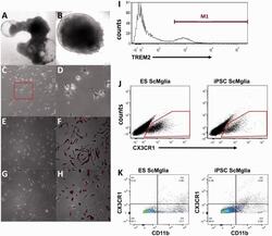
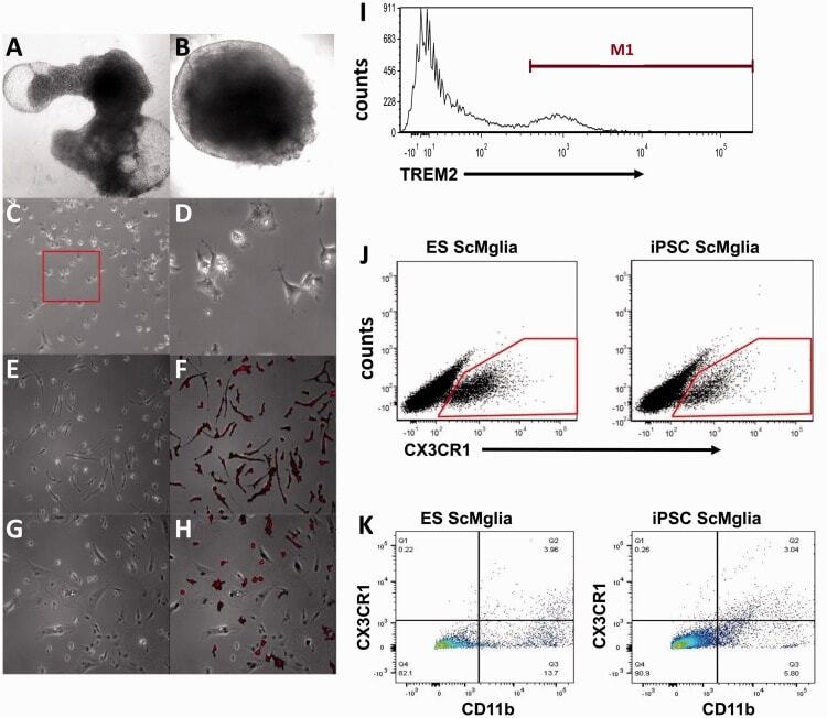
- Figure 2. Microglia-like cells (ScMglia) differentiated from hPSC develop microglia-like morphology and express microglia/myeloid markers. (a, b) Representative images (taken with 4 x objective) of cystic (a) and dense, noncystic (b) EBs in culture at Day 10. (c) Representative phase contrast images at 10 x of differentiating cells at Day 21 and (d) magnified 32 x images of microglial-like precursor cells at Day 21 exhibiting rounded morphologies with microglia-like processes (red box inset in (c)). (e) Phase image (32 x objective) of microglia-like cells (ScMglia) differentiated from iPS cells at Day 28, showing ramified morphology and even spacing among cells that have migrated away from the EB. (f) Most cells with this morphology also stained positively for microglial marker Iba1 (red). (g) Phase imaging showing fewer ScMglia were observed after extended culture (8 to 10 days after P1 plating) and (h) Iba1 immunolabeling demonstrates iPS-derived microglia-like cells continued to be Iba1 immunoreactive. Some microglia-like cells began to exhibit a more rounded morphology. (i) Representative flow cytometry analysis of ScMglia derived from an iPSC line showing 20.02% of gated cells express TREM2 (the M1 population); a total of 4,924 cells (events) were counted. (j) Flow cytometry analysis of ScMglia derived from both iPS and ES cells demonstrates both ES and iPSC-derived microglia-like cells express CX3CR1. (k) Differentiated ScMglia show double immunolabeling for CD11b and C
- Submitted by
- Invitrogen Antibodies (provider)
- Main image

- Experimental details
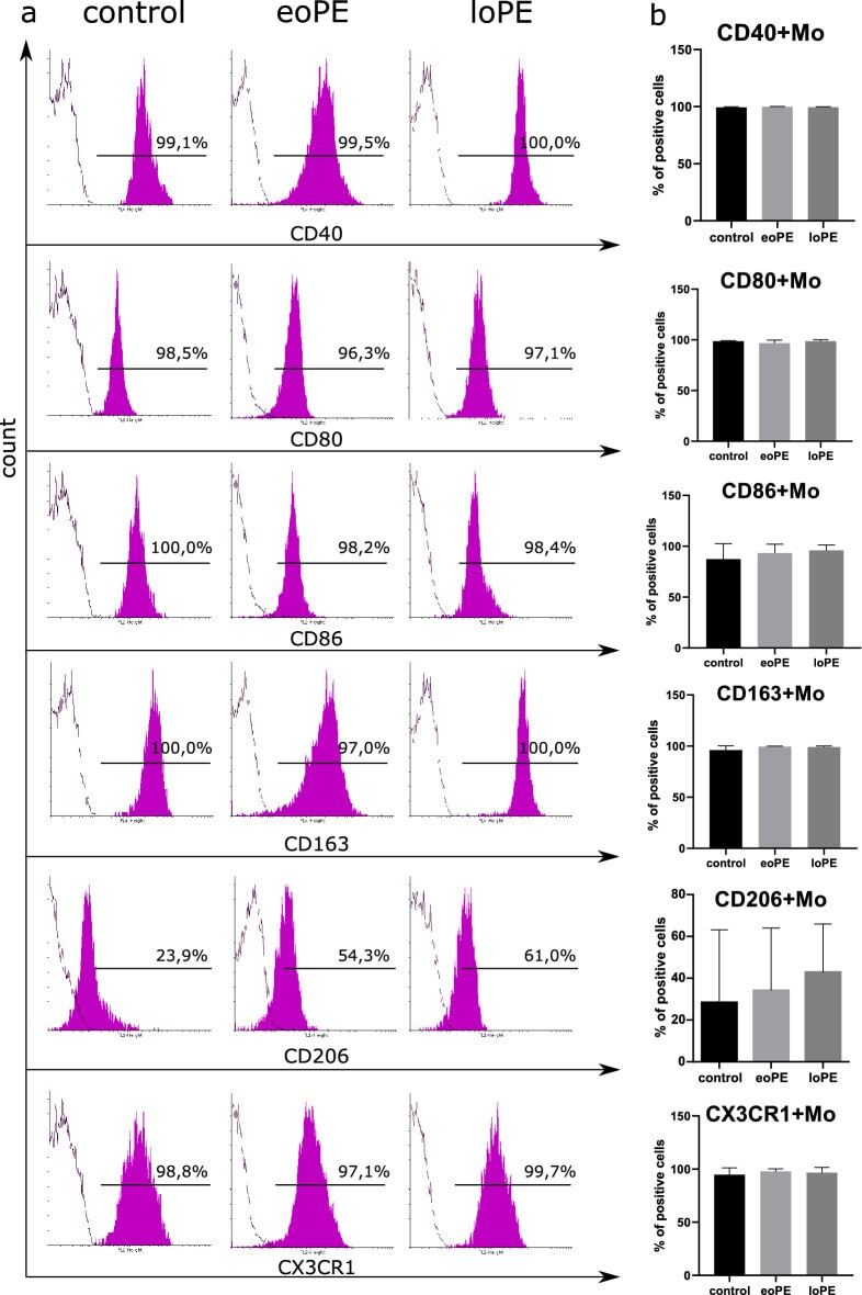
- Immunophenotyping of the patient's monocytes. Representative histograms of gated monocytes stained with antibodies to CD40, CD80, CD86, CD163, CD206 or CX3CR1 (magenta-filled histogram) and control samples (empty contour) for three groups of patients ( a ). Percents of positively stained cells are indicated above the gate bars. Levels of CD40+, CD80+, CD86+, CD163+, CD206+ and CX3CR1+ monocytes in three groups: the data are listed as mean +- SD ( b ): eoPE early-onset PE (n = 12), loPE late-onset PE (n = 10), control (n = 11).
- Submitted by
- Invitrogen Antibodies (provider)
- Main image
- Experimental details
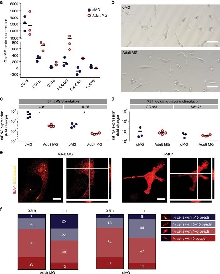
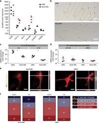
- Fig. 4 oMG expressed microglia-characteristic cell surface markers and showed similar functional immune and phagocytic properties as adult MG. a Flow cytometric analyses of the expression pattern of microglial extracellular markers on CD11b+-gated oMG (oMG 1, 3, and 5) compared to adult MG derived from three separate brain regions from adult MG1.1. (eight organoids were pooled per donor (oMG 1, 3, and 5) after 52 days in culture). b Morphology of magnetic automated cell sorted CD11b+ oMG 1 and adult MG in bright field microscope after 1 week in culture. Scale bar 40 mum. c mRNA expression, determined by qRT-PCR, of pro-inflammatory cytokines IL6 and IL1B after 6 h stimulation with LPS was significantly higher in oMG compared to adult MG (Mann-Whitney test IL6 and IL1B: U = 0, n = 4, p = 0.03). LPS-stimulated response relative to control condition without LPS. ( n = 4 experiments, eight organoids pooled per experiment; adult MG1.1) (* p < 0.05). d Anti-inflammatory response of oMG and adult MG was compared by qRT-PCR for expression of anti-inflammatory genes CD163 and MRC1 upon 72 h stimulation with dexamethasone. Dexamethasone-stimulated response relative to control condition without dexamethasone. (oMG, n = 3 separate experiments in which oMG were isolated from > 4 pooled cerebral organoids from iPSC 1 per experiment; adult MG, n = 4). e Phagocytosis capacity was tested oMG 1 and adult MG by performing a phagocytosis assay with iC3b-coated green-yellow