Explore
 Validate
 Learn
LearnNBP2-22471
antibody from Novus Biologicals
Targeting: STAT3
APRF
 Western blot
 Immunocytochemistry Immunoprecipitation Immunohistochemistry Chromatin Immunoprecipitation
Antibody data
- Antibody Data
- Antigen structure
- References [2]
- Comments [0]
- Validations
- Western blot [4]
- Immunoprecipitation [1]
- Immunohistochemistry [1]
- Chromatin Immunoprecipitation [1]
Submit
Validation data
Reference
Comment
Report error
- Product number
- NBP2-22471 - Provider product page

- Provider
- Novus Biologicals
- Product name
- Mouse Monoclonal STAT3 Antibody
- Antibody type
- Monoclonal
- Description
- Protein A purified.
- Reactivity
- Human, Mouse, Rat, Simian
- Host
- Mouse
- Isotype
- IgG
- Vial size
- 100 uL
- Concentration
- 1 mg/ml
- Storage
- Store at -20C. Avoid freeze-thaw cycles.
Submitted references STAT3, STAT5A, STAT5B and STAT6 proteins are overexpressed in human basal cell carcinoma.
Extracellular vesicles secreted by hypoxia pre-challenged mesenchymal stem cells promote non-small cell lung cancer cell growth and mobility as well as macrophage M2 polarization via miR-21-5p delivery.
Sławińska M, Lakomy J, Biernat W, Sokołowska-Wojdyło M, Karczewska J, Zabłotna M, Jankau J, Nowicki RJ, Sobjanek M
Clinical and experimental dermatology 2020 Mar;45(2):165-171
Clinical and experimental dermatology 2020 Mar;45(2):165-171
Extracellular vesicles secreted by hypoxia pre-challenged mesenchymal stem cells promote non-small cell lung cancer cell growth and mobility as well as macrophage M2 polarization via miR-21-5p delivery.
Ren W, Hou J, Yang C, Wang H, Wu S, Wu Y, Zhao X, Lu C
Journal of experimental & clinical cancer research : CR 2019 Feb 8;38(1):62
Journal of experimental & clinical cancer research : CR 2019 Feb 8;38(1):62
No comments: Submit comment
Supportive validation
- Submitted by
- Novus Biologicals (provider)
- Main image
- Experimental details
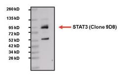
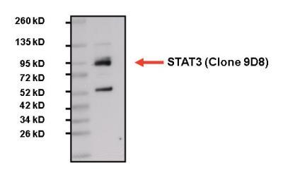
- Western Blot: STAT3 Antibody (9D8) [NBP2-22471] - Analysis of 25ug HepG2 total lysate.
- Submitted by
- Novus Biologicals (provider)
- Main image
- Experimental details
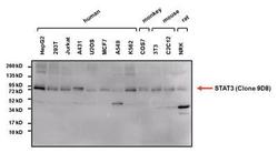
- Western Blot: STAT3 Antibody (9D8) [NBP2-22471] - Analysis of 25ug of various whole cell lysates.

- Submitted by
- Novus Biologicals (provider)
- Main image

- Experimental details
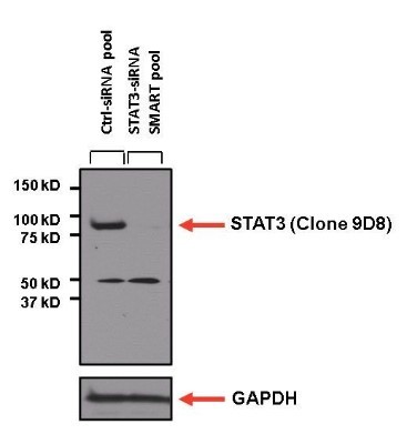
- Western Blot: STAT3 Antibody (9D8) [NBP2-22471] - Analysis of 25ug of U2-OS lysate from STAT3 SMART pool siRNA transfected or non-targeting control transfected U2-OS cells onto a 4-20% Tris-HCl polyacrylamide gel.
- Submitted by
- Novus Biologicals (provider)
- Main image
- Experimental details
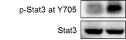
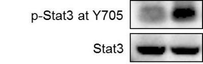
- Western Blot: STAT3 Antibody (9D8) [NBP2-22471] - Human breast cancer cell MDA-MB-231 was treated with carboplatin for 72 hours and the expression of p-Stat3 at Y705 and total Stat3 were detected by western blot. From verified customer review.
Supportive validation
- Submitted by
- Novus Biologicals (provider)
- Main image
- Experimental details
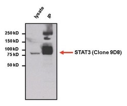
- Immunoprecipitation: STAT3 Antibody (9D8) [NBP2-22471] - Analysis of STAT3 was performed on HepG2 cells. The antigen: antibody complex was formed by incubating 750ug whole cell lysate with 2ug of mouse monoclonal antibody recognizing STAT3 overnight on a rocking platform at 4C. The immune-complex was then captured on 50ul Protein A/G Plus Agarose. Captured immune-complexes were then washed extensively and proteins eluted with 5X Reducing Sample Loading Dye. Samples were then resolved on a 4-20% Tris-HCl polyacrylamide gel. Proteins were transferred to PVDF membrane and blocked with 5% Milk/TBS-0.1%Tween for at least 1 hour. Membranes were then probed with a mouse monoclonal antibody recognizing STAT3 at a dilution of 1:5000 overnight rotating at 4C. Membranes were washed in TBST and probed with Clean-blot IP detection reagent at a dilution of 1:2000 for at least one hour. Membranes were washed and chemiluminescent detection was performed using Super Signal West Dura.

Supportive validation
- Submitted by
- Novus Biologicals (provider)
- Main image
- Experimental details
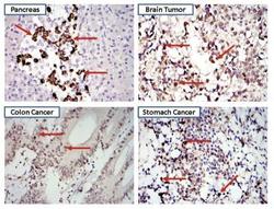
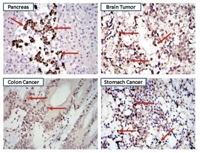
- Immunohistochemistry-Paraffin: STAT3 Antibody (9D8) [NBP2-22471] - Biopsies of normal and cancer tissues.
Supportive validation
- Submitted by
- Novus Biologicals (provider)
- Main image
- Experimental details
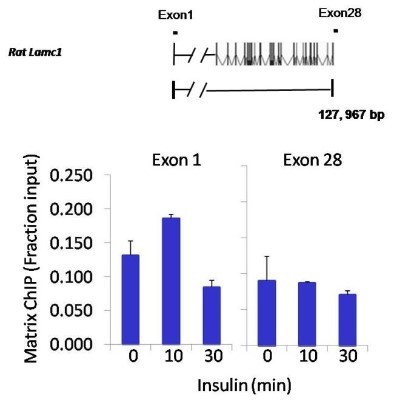
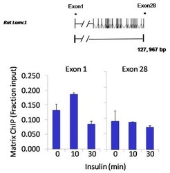
- Chromatin Immunoprecipitation: STAT3 Antibody (9D8) [NBP2-22471] - Analysis performed using cross-linked chromatin from rat hepatoma cells treated with insulin. IP performed using a multiplex microplate Matrix ChIP assay with STAT3 monoclonal antibody. Chromatin aliquots from cells were used per ChIP pull-down. Quantitative PCR data done in quadruplicate using 1ul of DNA in 2ul SYBR real-time PCR reactions containing primers to amplify exon-1 or exon-28 of LAMC1.Quantitation of immunoprecipitated chromatin is presented as signal relative to the total amount of input chromatin. Results represent the mean +/- SEM for three experiments. A schematic representation of the rat LAMC1 locus is shown; boxes represent exons (black boxes = translated regions, white boxes = untranslated regions), the zigzag line represents an intron, and the straight line represents upstream sequence. Regions amplified by LAMC1 primers are represented by black bars. Data courtesy of the Innovators Program.