Explore
 Validate
 Learn
LearnGTX15789
antibody from GeneTex
Targeting: STAT3
APRF
 Western blot Immunocytochemistry Immunoprecipitation
 Immunohistochemistry Chromatin Immunoprecipitation
Antibody data
- Antibody Data
- Antigen structure
- References [1]
- Comments [0]
- Validations
- Western blot [1]
- Immunocytochemistry [1]
- Immunoprecipitation [1]
Submit
Validation data
Reference
Comment
Report error
- Product number
- GTX15789 - Provider product page

- Provider
- GeneTex
- Product name
- STAT3 antibody [9D8]
- Antibody type
- Monoclonal
- Reactivity
- Human, Mouse, Rat, Simian
- Host
- Mouse
Submitted references Targeted regulationof STAT3 by miR-29a in mediating Taxol resistance of nasopharyngeal carcinoma cell line CNE-1.
Gao J, Shao Z, Yan M, Fu T, Zhang L, Yan Y
Cancer biomarkers : section A of Disease markers 2018;22(4):641-648
Cancer biomarkers : section A of Disease markers 2018;22(4):641-648
No comments: Submit comment
Supportive validation
- Submitted by
- GeneTex (provider)
- Main image
- Experimental details
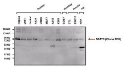
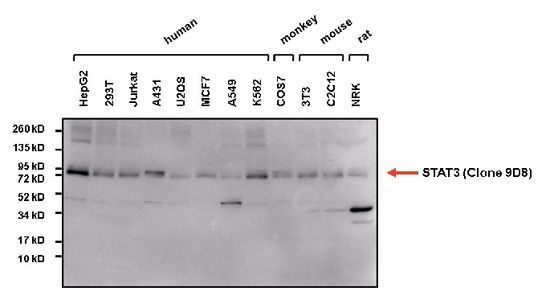
- Western blot analysis of STAT3 in 25ug of various whole cell lysates. Proteins were transferred to a PVDF membrane and blocked with 5% Milk/TBST for at least 1 hour. Membranes were probed with STAT3 antibody [9D8] at a dilution of 1:5000 overnight at 4¢XC on a rocking platform. Membranes were washed in TBS-0.1%Tween 20 and probed with a HRP-conjugated secondary antibody. Membranes were washed and chemiluminescent detection was performed.
Supportive validation
- Submitted by
- GeneTex (provider)
- Main image
- Experimental details
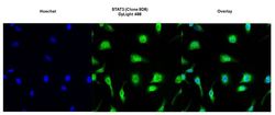
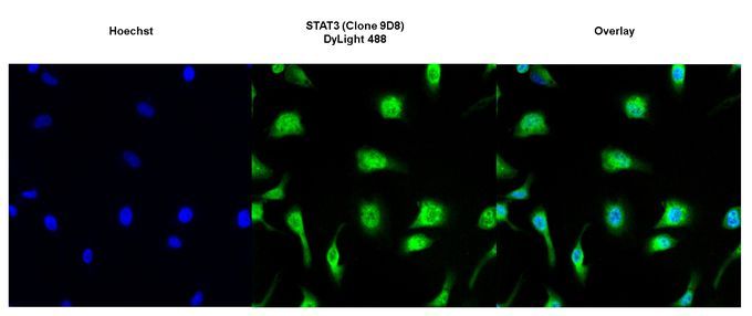
- Immunofluorescent analysis of STAT3 (green) in HeLa cells. Formalin-fixed cells were permeabilized with 0.1% Triton X-100 in TBS for 10 minutes at room temperature. Cells were then blocked with 1% BSA for 15 minutes at room temperature. Cells were probed with STAT3 antibody [9D8] at a dilution of 1:100 for at least 1 hour at room temperature. Cells were then washed with PBS and incubated with DyLight 488-conjugated secondary antibody. Nuclei (blue) were stained with Hoechst 33342 dye. Images were taken at 20X magnification.
Supportive validation
- Submitted by
- GeneTex (provider)
- Main image
- Experimental details
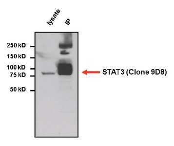
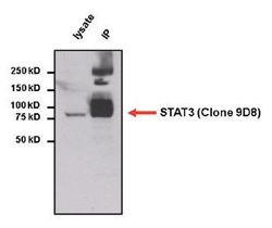
- Immunoprecipitation of STAT3 from HepG2 cell lysate. The antigen-antibody complex was formed by incubating 750 £gg of whole cell lysate with 2 £gg of STAT3 antibody [9D8] overnight on a rocking platform at 4¢XC. The immune-complex was then captured on 50 £gl Protein A/G Agarose. Captured immune-complexes were then washed extensively and eluted. Samples were then resolved on a 4-20% Tris-HCl polyacrylamide gel. Proteins were transferred to PVDF membrane and blocked with 5% Milk/TBS-0.1%Tween for at least 1 hour. Membranes were then probed with STAT3 antibody [9D8] at a dilution of 1:5000 overnight rotating at 4¢XC. Membranes were washed in TBST and probed with a proper secondary antibody. Membranes were washed and chemiluminescent detection was performed.