Explore
 Validate
 Learn
Learn Western blot
 ELISA
ELISAAntibody data
- Antibody Data
- Antigen structure
- References [2]
- Comments [0]
- Validations
- Western blot [1]
- Immunocytochemistry [2]
- Immunohistochemistry [3]
- Chromatin Immunoprecipitation [2]
- Other assay [3]
Submit
Validation data
Reference
Comment
Report error
- Product number
- 710077 - Provider product page

- Provider
- Invitrogen Antibodies
- Product name
- STAT3 Recombinant Polyclonal Antibody
- Antibody type
- Polyclonal
- Antigen
- Recombinant full-length protein
- Reactivity
- Human, Mouse
- Host
- Rabbit
- Isotype
- IgG
- Vial size
- 100 µg
- Concentration
- 0.5 mg/mL
- Storage
- Store at 4°C short term. For long term storage, store at -20°C, avoiding freeze/thaw cycles.
Submitted references Krüppel-like factor 7 attenuates hippocampal neuronal injury after traumatic brain injury.
Role and mechanism of nursing cooperation and tetramethylpyrazine application in post-operative pain in patients undergoing total knee arthroplasty.
Li WY, Fu XM, Wang ZD, Li ZG, Ma D, Sun P, Liu GB, Zhu XF, Wang Y
Neural regeneration research 2022 Mar;17(3):661-672
Neural regeneration research 2022 Mar;17(3):661-672
Role and mechanism of nursing cooperation and tetramethylpyrazine application in post-operative pain in patients undergoing total knee arthroplasty.
Liu C, Liu R, Tang M, Yang X, Gong X
Experimental and therapeutic medicine 2019 Mar;17(3):2366-2372
Experimental and therapeutic medicine 2019 Mar;17(3):2366-2372
No comments: Submit comment
Supportive validation
- Submitted by
- Invitrogen Antibodies (provider)
- Main image

- Experimental details
- Western blot analysis of STAT3 in whole cell extract (80 µg) from HeLa cells treated with IFN1a (150 ng/mL, 15 min) using a STAT3 Recombinant Rabbit Polyclonal Antibody (Product # 710077) at a dilution of 5 µg/mL. Detection was performed using an HRP-conjugated Goat anti-Rabbit secondary antibody at a dilution of 1:5000 followed by chemiluminescence (ECL). Results show a band at ~84 kDa.

Supportive validation
- Submitted by
- Invitrogen Antibodies (provider)
- Main image

- Experimental details
- Immunofluorescence analysis of STAT3 was done on 70% confluent log phase MCF-7 cells. The cells were fixed with 4% paraformaldehyde for 10 minutes, permeabilized with 0.1% Triton™ X-100 for 10 minutes, and blocked with 1% BSA for 1 hour at room temperature. The cells were labeled with STAT3 Antibody (3HCLC), Recombinant Rabbit Polyclonal Antibody (Product # 710077) at 1 µg/mL in 0.1% BSA and incubated for 3 hours at room temperature and then labeled with Goat anti-Rabbit IgG (H+L) Superclonal™ Secondary Antibody, Alexa Fluor® 488 conjugate (Product # A27034) at a dilution of 1:2000 for 45 minutes at room temperature (Panel a: green). Nuclei (Panel b: blue) were stained with SlowFade® Gold Antifade Mountant with DAPI (Product # S36938). F-actin (Panel c: red) was stained with Alexa Fluor® 555 Rhodamine Phalloidin (Product # R415, 1:300). Panel d is a merged image showing Nuclear and Cytoplasmic localization. Panel e is a no primary antibody control. The images were captured at 60X magnification.

- Submitted by
- Invitrogen Antibodies (provider)
- Main image

- Experimental details
- Immunofluorescence analysis of STAT3 was performed using 70 % confluent log phase HeLa cells. The cells were fixed with 4% paraformaldehyde for 10 minutes, permeabilized with 0.1% Triton™ X-100 for 10 minutes, and blocked with 1% BSA for 1 hour at room temperature. The cells were labeled with STAT3 Recombinant Rabbit Polyclonal Antibody (Product # 710077 ) at 5µg/mL in 0.1% BSA and incubated for 3 hours at room temperature and then labeled with Goat anti-Rabbit IgG (H+L) Superclonal™ Secondary Antibody, Alexa Fluor® 488 conjugate (A27034) at a dilution of 1:2000 for 45 minutes at room temperature (Panel a: green). Nuclei (Panel b: blue) were stained with SlowFade® Gold Antifade Mountant with DAPI (Product # S36938). F-actin (Panel c: red) was stained with Rhodamine Phalloidin (Product # R415, 1:300). Panel d represents the merged image showing Cytoplasmic and Nuclear localization. Panel e shows the no primary antibody control. The images were captured at 60X magnification.

Supportive validation
- Submitted by
- Invitrogen Antibodies (provider)
- Main image
- Experimental details
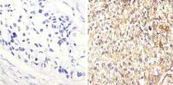
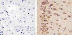
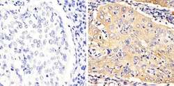
- Immunohistochemistry analysis of STAT3 showing staining in the cytoplasm of paraffin-embedded human lung squamous carcinoma (right) compared to a negative control without primary antibody (left). To expose target proteins, antigen retrieval was performed using 10mM sodium citrate (pH 6.0), microwaved for 8-15 min. Following antigen retrieval, tissues were blocked in 3% H2O2-methanol for 15 min at room temperature, washed with ddH2O and PBS, and then probed with a STAT3 Recombinant Rabbit Polyclonal Antibody (clone 3HCLC) (Product # 710077) diluted in 3% BSA-PBS at a dilution of 1:20 overnight at 4°C in a humidified chamber. Tissues were washed extensively in PBST and detection was performed using an HRP-conjugated secondary antibody followed by colorimetric detection using a DAB kit. Tissues were counterstained with hematoxylin and dehydrated with ethanol and xylene to prep for mounting.

- Submitted by
- Invitrogen Antibodies (provider)
- Main image
- Experimental details
- Immunohistochemistry analysis of STAT3 showing staining in the cytoplasm of paraffin-embedded mouse brain tissue (right) compared to a negative control without primary antibody (left). To expose target proteins, antigen retrieval was performed using 10mM sodium citrate (pH 6.0), microwaved for 8-15 min. Following antigen retrieval, tissues were blocked in 3% H2O2-methanol for 15 min at room temperature, washed with ddH2O and PBS, and then probed with a STAT3 Recombinant Rabbit Polyclonal Antibody (clone 3HCLC) (Product # 710077) diluted in 3% BSA-PBS at a dilution of 1:20 overnight at 4°C in a humidified chamber. Tissues were washed extensively in PBST and detection was performed using an HRP-conjugated secondary antibody followed by colorimetric detection using a DAB kit. Tissues were counterstained with hematoxylin and dehydrated with ethanol and xylene to prep for mounting.

- Submitted by
- Invitrogen Antibodies (provider)
- Main image
- Experimental details
- Immunohistochemistry analysis of STAT3 showing staining in the cytoplasm of paraffin-embedded human cervical carcinoma (right) compared to a negative control without primary antibody (left). To expose target proteins, antigen retrieval was performed using 10mM sodium citrate (pH 6.0), microwaved for 8-15 min. Following antigen retrieval, tissues were blocked in 3% H2O2-methanol for 15 min at room temperature, washed with ddH2O and PBS, and then probed with a STAT3 Recombinant Rabbit Polyclonal Antibody (clone 3HCLC) (Product # 710077) diluted in 3% BSA-PBS at a dilution of 1:20 overnight at 4°C in a humidified chamber. Tissues were washed extensively in PBST and detection was performed using an HRP-conjugated secondary antibody followed by colorimetric detection using a DAB kit. Tissues were counterstained with hematoxylin and dehydrated with ethanol and xylene to prep for mounting.

Supportive validation
- Submitted by
- Invitrogen Antibodies (provider)
- Main image

- Experimental details
- Enrichment of endogenous STAT3 protein at specific gene loci using Anti-STAT3 Recombinant Rabbit Polyclonal Antibody: Chromatin Immunoprecipitation (ChIP) was performed using Anti-STAT3 Recombinant Rabbit Polyclonal Antibody (Product # 710077, 5 µg) on sheared chromatin from 2 million HeLa cells treated with 100 ng/mL of IFN alpha for 30 minutes using the "MAGnify ChIP system" kit (Product # 49-2024). Normal Rabbit IgG was used as a negative IP control. The purified DNA was analyzed by 7500 Fast qPCR system (Product # 4351106) with optimized PCR primer pairs for the promoter of active GLS1, OAS1, cFOS gene, used as positive control target, and the SAT2, used as negative control target. Data is presented as fold enrichment of the antibody signal versus the negative control IgG using the comparative CT method.

- Submitted by
- Invitrogen Antibodies (provider)
- Main image

- Experimental details
- Enrichment of endogenous STAT3 protein at specific gene loci using Anti-STAT3 Recombinant Rabbit Polyclonal Antibody: Chromatin Immunoprecipitation (ChIP) was performed using Anti-STAT3 Recombinant Rabbit Polyclonal Antibody (Product # 710077, 5 µg) on sheared chromatin from 2 million HeLa cells and 2 million HeLa cells treated with 100 ng/mL of IFN alpha for 30 minutes using the MAGnify ChIP system kit (Product # 49-2024). Normal Rabbit IgG was used as a negative IP control. The purified DNA was analyzed by 7500 Fast qPCR system (Product # 4351106) with optimized PCR primer pairs for the promoter of active GLS1, AP1 gene, used as positive control target, and the MYOD, SAT2, used as negative control target. Data is presented as fold enrichment of the antibody signal versus the negative control IgG using the comparative CT method.

Supportive validation
- Submitted by
- Invitrogen Antibodies (provider)
- Main image

- Experimental details
- Indirect ELISA analysis of endogenous STAT3 in HeLa cells treated with IFN1a (150 ng/mL for 15 min) and whole cell lysate (1 µg/well) using a STAT3 Recombinant Rabbit Polyclonal Antibody (Product # 710077) followed by detection using an HRP-conjugated Goat anti-Rabbit secondary antibody (Product # G-21234) and TMB (Product # SB01) as a substrate.

- Submitted by
- Invitrogen Antibodies (provider)
- Main image
- Experimental details
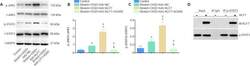
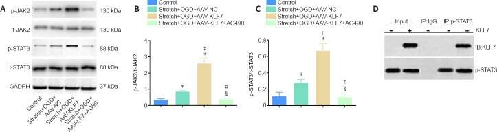
- Figure 4 Involvement of the JAK2/STAT3 signaling pathway in the neuroprotective effects of KLF7. (A) Representative western blot of p-JAK2, t-JAK2, p-STAT3, and t-STAT3 expression in cultured HT22 cells at 1 day after stretch and OGD treatment. (B, C) Quantitative results of the relative optical densities of p-JAK2/t-JAK2 and p-STAT3/t-STAT3. (D) Co-immunoprecipitation analysis of the interactions between KLF7 and p-STAT3 in stretch- and OGD-damaged HT22 cells at 1 day, revealing a physical interaction between KLF7 and p-STAT3. Data are expressed as the mean +- SD. Each experiment was repeated three times. * P < 0.05, vs . control group; # P < 0.05, vs . stretch + OGD + AAV-NC group; & P < 0.05, vs . stretch + OGD + AAV-KLF7 group (one-way analysis of variance followed by Tukey's post hoc test). AAV: Adeno-associated virus; GAPDH: glyceraldehyde-3-phosphate dehydrogenase; IB: immunoblotting; IP: immunoprecipitation; KLF7: Kruppel-like factor 7; NC: negative control; OGD: oxygen-glucose deprivation; p-JAK2: phospho-Janus kinase 2; t-JAK2: total-Janus kinase 2; p-STAT3: phospho-signal transducer and activator of transcription 3; t-STAT3: total-signal transducer and activator of transcription 3.
- Submitted by
- Invitrogen Antibodies (provider)
- Main image
- Experimental details
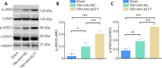
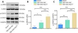
- Figure 9 KLF7 activates the JAK2/STAT3 pathway at 3 days after TBI. (A) Representative western blots of p-JAK2, t-JAK2, p-STAT3, and t-STAT3 expression in ipsilateral hippocampal tissue. (B, C) Quantitative results of the p-JAK2/t-JAK2 and p-STAT3/t-STAT3 ratios. Data are expressed as the mean +- SD ( n = 3). * P < 0.05, ** P < 0.01, *** P < 0.001 (one-way analysis of variance followed by Tukey's post hoc test). AAV: Adeno-associated virus; GAPDH: glyceraldehyde-3-phosphate dehydrogenase; KLF7: Kruppel-like factor 7; NC: negative control; p-JAK2: phospho-Janus kinase 22; p-STAT3: phospho-signal transducer and activator of transcription 3; t-JAK2: total-Janus kinase; t-STAT3: total-signal transducer and activator of transcription 3; TBI: traumatic brain injury.