 Explore
 Validate
 Learn
Learn Western blot
Antibody data
- Antibody Data
- Antigen structure
- References [3]
- Comments [0]
- Validations
- Western blot [3]
- Immunohistochemistry [1]
- Chromatin Immunoprecipitation [1]
Submit
Validation data
Reference
Comment
Report error
- Product number
- 710093 - Provider product page

- Provider
- Invitrogen Antibodies
- Product name
- Phospho-STAT3 (Tyr705) Recombinant Polyclonal Antibody (3HCLC)
- Antibody type
- Polyclonal
- Antigen
- Synthetic peptide
- Description
- This antibody is predicted to react with mouse, rat and non-human primate based on sequence homology.
- Antibody clone number
- 3HCLC
- Concentration
- 0.5 mg/mL
Submitted references Recombinant human erythropoietin and interferon-β-1b protect against 3-nitropropionic acid-induced neurotoxicity in rats: possible role of JAK/STAT signaling pathway.
Activation of Interferon Signaling in Chronic Lymphocytic Leukemia Cells Contributes to Apoptosis Resistance via a JAK-Src/STAT3/Mcl-1 Signaling Pathway.
Relation of Neutrophil Gelatinase-Associated Lipocalin Overexpression to the Resistance to Apoptosis of Tumor B Cells in Chronic Lymphocytic Leukemia.
Sayed RH, Ghazy AH, Yammany MFE
Inflammopharmacology 2022 Apr;30(2):667-681
Inflammopharmacology 2022 Apr;30(2):667-681
Activation of Interferon Signaling in Chronic Lymphocytic Leukemia Cells Contributes to Apoptosis Resistance via a JAK-Src/STAT3/Mcl-1 Signaling Pathway.
Bauvois B, Pramil E, Jondreville L, Quiney C, Nguyen-Khac F, Susin SA
Biomedicines 2021 Feb 13;9(2)
Biomedicines 2021 Feb 13;9(2)
Relation of Neutrophil Gelatinase-Associated Lipocalin Overexpression to the Resistance to Apoptosis of Tumor B Cells in Chronic Lymphocytic Leukemia.
Bauvois B, Pramil E, Jondreville L, Chapiro E, Quiney C, Maloum K, Susin SA, Nguyen-Khac F
Cancers 2020 Jul 31;12(8)
Cancers 2020 Jul 31;12(8)
No comments: Submit comment
Supportive validation
- Submitted by
- Invitrogen Antibodies (provider)
- Main image

- Experimental details
- Western blot analysis of Phospho-STAT3 pTyr705 in whole cell extracts of HeLa cells treated with 100 ng/mL insulin for 15 min (lane1), without insulin treatment (lane 2), and treated with 150 ng/mL IFN1a for 15 min (lane 3) using a Phospho-STAT3 pTyr705 Recombinant Rabbit Polyclonal Antibody (Product # 710093) at a dilution of 2.5 µg/mL.

- Submitted by
- Invitrogen Antibodies (provider)
- Main image
- Experimental details
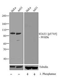
- Western blot analysis of STAT3 (pY705) was performed by loading 20 µg of HeLa (lane1) and A431 (lane2) cell lysates using Novex®NuPAGE®4-12 % Bis-Tris gel (Product # NP0321BOX), XCell SureLock Electrophoresis System (Product # EI0002), Novex® Sharp Pre-Stained Protein Standard (Product # LC5800). Proteins were transferred to a PVDF membrane and blocked with 5 % skim milk for 1 hour at room temperature. STAT3 (pY705) was detected at ~90 kDa using STAT3 (pY705) Recombinant Rabbit Polyclonal Antibody (Product # 710093) at 1 µg-3 µg/mL in 2.5 % skim milk at 4°C overnight on a rocking platform. To confirm specificity, the corresponding blot on right was incubated with Lambda phosphatase and its reactivity with antibody was tested. Goat anti-Rabbit IgG-HRP Secondary Antibody (Product # G-21234) at 1:5000 dilution was used and chemiluminescent detection was performed using Pierce™ ECL Western blotting Substrate (Product # 32106).

- Submitted by
- Invitrogen Antibodies (provider)
- Main image

- Experimental details
- Western blot analysis of Phospho-STAT3 pTyr705 in whole cell extracts of HeLa cells treated with 100 ng/mL insulin for 15 min (lane1), without insulin treatment (lane 2), and treated with 150 ng/mL IFN1a for 15 min (lane 3) using a Phospho-STAT3 pTyr705 Recombinant Rabbit Polyclonal Antibody (Product # 710093) at a dilution of 2.5 µg/mL.

Supportive validation
- Submitted by
- Invitrogen Antibodies (provider)
- Main image

- Experimental details
- Immunohistochemistry analysis of STAT3 (pY705) showing staining in the cytoplasm of paraffin-embedded human cervical carcinoma (right) compared to a negative control without primary antibody (left). To expose target proteins, antigen retrieval was performed using 10mM sodium citrate (pH 6.0), microwaved for 8-15 min. Following antigen retrieval, tissues were blocked in 3% H2O2-methanol for 15 min at room temperature, washed with ddH2O and PBS, and then probed with a STAT3 (pY705) Recombinant Rabbit Polyclonal Antibody (clone 3HCLC) (Product # 710093) diluted in 3% BSA-PBS at a dilution of 1:20 overnight at 4°C in a humidified chamber. Tissues were washed extensively in PBST and detection was performed using an HRP-conjugated secondary antibody followed by colorimetric detection using a DAB kit. Tissues were counterstained with hematoxylin and dehydrated with ethanol and xylene to prep for mounting.

Supportive validation
- Submitted by
- Invitrogen Antibodies (provider)
- Main image

- Experimental details
- ChIP- qPCR analysis of STAT3 (pY705) was performed with 3 µg/mL of the STAT3 (pY705) Recombinant Rabbit Polyclonal Antibody (Product # 710093) on sheared chromatin from 2 million HeLa cells treated with IFN-alpha (50 ng/mL) for 1h using the MAGnify Chromatin Immunoprecipitation System (Product # 49-2024). Normal rabbit IgG (3 µg/mL) was used as a negative IP control. The purified DNA from each ChIP sample was analyzed by StepOnePlus Real-Time PCR System (Product # 4376600) with primers for the promoter of active GLS1 gene, used as positive control target, and the OAS1 gene, used as negative control target. Data is presented as fold enrichment of the antibody signal versus the negative control IgG using the comparative CT method.
