Explore
 Validate
 Learn
Learn Western blot
 Immunohistochemistry
Antibody data
- Antibody Data
- Antigen structure
- References [21]
- Comments [0]
- Validations
- Immunohistochemistry [1]
Submit
Validation data
Reference
Comment
Report error
- Product number
- P00007-2 - Provider product page

- Provider
- Boster Biological Technology
- Product name
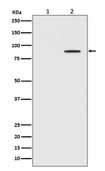
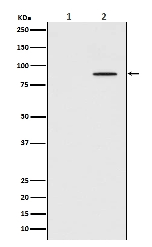
- Anti-Phospho-STAT3 (Y705) Rabbit Monoclonal Antibody
- Antibody type
- Monoclonal
- Description
- Monoclonal antibody for STAT3 detection. Host: Rabbit.Size: 100ug/vial. Tested applications: IP, IF, IHC, ICC, WB. Reactive species: Human, Mouse, Rat STAT3 information: Molecular Weight: 88068 MW; Subcellular Localization: Cytoplasm. Nucleus. Shuttles between the nucleus and the cytoplasm. Translocated into the nucleus upon tyrosine phosphorylation and dimerization, in response to signaling by activated FGFR1, FGFR2, FGFR3 or FGFR4. Constitutive nuclear presence is independent of tyrosine phosphorylation. Predominantly present in the cytoplasm without stimuli. Upon leukemia inhibitory factor (LIF) stimulation, accumulates in the nucleus. The complex composed of BART and ARL2 plays an important role in the nuclear translocation and retention of STAT3. Identified in a complex with LYN and PAG1; Tissue Specificity: Heart, brain, placenta, lung, liver, skeletal muscle, kidney and pancreas.
- Reactivity
- Human, Mouse, Rat
- Host
- Rabbit
- Antibody clone number
- IFA-19
- Vial size
- 100ug/vial
- Concentration
- 0.5-1mg/ml, actual concentration vary by lot. Use suggested dilution ratio to decide dilution procedure.
- Storage
- At -20°C for one year. Avoid repeated freezing and thawing.
Submitted references Pancreatic neuroendocrine tumor progression and resistance to everolimus: the crucial role of NF-kB and STAT3 interplay.
α-Linolenic acid ameliorates pentylenetetrazol-induced neuron apoptosis and neurological impairment in mice with seizures via down-regulating JAK2/STAT3 pathway.
Somatic PDGFRB activating variants promote smooth muscle cell phenotype modulation in intracranial fusiform aneurysm.
PCSK5 downregulation promotes the inhibitory effect of andrographolide on glioblastoma through regulating STAT3.
Modified Biejia Jianwan decoction restrains PD-L1-mediated immune evasion through the HIF-1α/STAT3/NF-κB signaling pathway.
The direct impact of pegvisomant on osteoblast functions and bone development.
Integration analysis using bioinformatics and experimental validation on the clinical and biological significance of TSLP in cancers.
(2E)-1-(2,4,6-Trimethoxyphenyl)-3-(4-chlorophenyl)prop-2-en-1-one, a Chalcone Derivative, Promotes Apoptosis by Suppressing RAS-ERK and AKT/FOXO3a Pathways in Hepatocellular Carcinoma Cells.
The Large Molecular Weight Polysaccharide from Wild Cordyceps and Its Antitumor Activity on H22 Tumor-Bearing Mice.
Curcumin and its nano-formulations: Defining triple-negative breast cancer targets through network pharmacology, molecular docking, and experimental verification.
Folic acid intervention changes liver Foxp3 methylation and ameliorates the damage caused by Th17/Treg imbalance after long-term alcohol exposure.
Cryptotanshinone ameliorates MPP(+)-induced oxidative stress and apoptosis of SH-SY5Y neuroblastoma cells: the role of STAT3 in Parkinson's disease.
Triptolide attenuates LPS-induced activation of RAW 264.7 macrophages by inducing M1-to-M2 repolarization via the mTOR/STAT3 signaling.
Overexpression of miR-125a-5p Inhibits Hepatocyte Proliferation through the STAT3 Regulation In Vivo and In Vitro.
The expression of Hexokinase 2 and its hub genes are correlated with the prognosis in glioma.
Water extract of sporoderm-broken spores of Ganoderma lucidum enhanced pd-l1 antibody efficiency through downregulation and relieved complications of pd-l1 monoclonal antibody.
JAK2/STAT3 regulates estrogen-related senescence of bone marrow stem cells.
JAK2-STAT3 signaling pathway is involved in rat periapical lesions induced by Enterococcus faecalis.
Glycosylation of dentin matrix protein 1 is critical for fracture healing via promoting chondrogenesis.
ELP2 negatively regulates osteoblastic differentiation impaired by tumor necrosis factor α in MC3T3-E1 cells through STAT3 activation.
Prognostic role of STAT3 in solid tumors: a systematic review and meta-analysis.
Vitali E, Valente G, Panzardi A, Laffi A, Zerbi A, Uccella S, Mazziotti G, Lania A
Journal of endocrinological investigation 2024 May;47(5):1101-1117
Journal of endocrinological investigation 2024 May;47(5):1101-1117
α-Linolenic acid ameliorates pentylenetetrazol-induced neuron apoptosis and neurological impairment in mice with seizures via down-regulating JAK2/STAT3 pathway.
Zeng X, Luo F, Cheng YH, Gao J, Hong D
The British journal of nutrition 2024 May 22;:1-12
The British journal of nutrition 2024 May 22;:1-12
Somatic PDGFRB activating variants promote smooth muscle cell phenotype modulation in intracranial fusiform aneurysm.
Hao L, Ya X, Wu J, Tao C, Ma R, Zheng Z, Mou S, Ling Y, Yang Y, Wang J, Zhang Y, Lin Q, Zhao J
Journal of biomedical science 2024 May 13;31(1):51
Journal of biomedical science 2024 May 13;31(1):51
PCSK5 downregulation promotes the inhibitory effect of andrographolide on glioblastoma through regulating STAT3.
Gong H, Yang X, An L, Zhang W, Liu X, Shu L, Yang L
Molecular and cellular biochemistry 2024 Mar 29;
Molecular and cellular biochemistry 2024 Mar 29;
Modified Biejia Jianwan decoction restrains PD-L1-mediated immune evasion through the HIF-1α/STAT3/NF-κB signaling pathway.
Tian X, Liu F, Wang Z, Zhang J, Liu Q, Zhang Y, Zhang D, Huang C, Zhao J, Jiang S
Journal of ethnopharmacology 2024 Mar 25;322:117577
Journal of ethnopharmacology 2024 Mar 25;322:117577
The direct impact of pegvisomant on osteoblast functions and bone development.
Vitali E, Grasso A, Schiavone ML, Trivellin G, Sobacchi C, Mione M, Mazziotti G, Lania A
Journal of endocrinological investigation 2024 Jun;47(6):1385-1394
Journal of endocrinological investigation 2024 Jun;47(6):1385-1394
Integration analysis using bioinformatics and experimental validation on the clinical and biological significance of TSLP in cancers.
Qu H, Liu X, Jiang T, Huang G, Cai H, Xing D, Mao Y, Zheng X
Cellular signalling 2023 Nov;111:110874
Cellular signalling 2023 Nov;111:110874
(2E)-1-(2,4,6-Trimethoxyphenyl)-3-(4-chlorophenyl)prop-2-en-1-one, a Chalcone Derivative, Promotes Apoptosis by Suppressing RAS-ERK and AKT/FOXO3a Pathways in Hepatocellular Carcinoma Cells.
Zhang M, Li J, Ma R, Xi J, Xi L, Zhang B, Tian J, Bai Z
Chemistry & biodiversity 2023 Jul;20(7):e202300050
Chemistry & biodiversity 2023 Jul;20(7):e202300050
The Large Molecular Weight Polysaccharide from Wild Cordyceps and Its Antitumor Activity on H22 Tumor-Bearing Mice.
Tan L, Liu S, Li X, He J, He L, Li Y, Yang C, Li Y, Hua Y, Guo J
Molecules (Basel, Switzerland) 2023 Apr 10;28(8)
Molecules (Basel, Switzerland) 2023 Apr 10;28(8)
Curcumin and its nano-formulations: Defining triple-negative breast cancer targets through network pharmacology, molecular docking, and experimental verification.
Deng Z, Chen G, Shi Y, Lin Y, Ou J, Zhu H, Wu J, Li G, Lv L
Frontiers in pharmacology 2022;13:920514
Frontiers in pharmacology 2022;13:920514
Folic acid intervention changes liver Foxp3 methylation and ameliorates the damage caused by Th17/Treg imbalance after long-term alcohol exposure.
Zhao H, Guo P, Zuo Y, Wang Y, Zhao H, Lan T, Xue M, Zhang H, Liang H
Food & function 2022 May 10;13(9):5262-5274
Food & function 2022 May 10;13(9):5262-5274
Cryptotanshinone ameliorates MPP(+)-induced oxidative stress and apoptosis of SH-SY5Y neuroblastoma cells: the role of STAT3 in Parkinson's disease.
Wang Q, Liu Y
Metabolic brain disease 2022 Jun;37(5):1477-1485
Metabolic brain disease 2022 Jun;37(5):1477-1485
Triptolide attenuates LPS-induced activation of RAW 264.7 macrophages by inducing M1-to-M2 repolarization via the mTOR/STAT3 signaling.
Zhu H, Tong S, Yan C, Zhou A, Wang M, Li C
Immunopharmacology and immunotoxicology 2022 Dec;44(6):894-901
Immunopharmacology and immunotoxicology 2022 Dec;44(6):894-901
Overexpression of miR-125a-5p Inhibits Hepatocyte Proliferation through the STAT3 Regulation In Vivo and In Vitro.
Zhang C, Zhao Y, Wang Q, Qin J, Ye B, Xu C, Yu G
International journal of molecular sciences 2022 Aug 4;23(15)
International journal of molecular sciences 2022 Aug 4;23(15)
The expression of Hexokinase 2 and its hub genes are correlated with the prognosis in glioma.
Huang Y, Ouyang F, Yang F, Zhang N, Zhao W, Xu H, Yang X
BMC cancer 2022 Aug 18;22(1):900
BMC cancer 2022 Aug 18;22(1):900
Water extract of sporoderm-broken spores of Ganoderma lucidum enhanced pd-l1 antibody efficiency through downregulation and relieved complications of pd-l1 monoclonal antibody.
He J, Zhang W, Di T, Meng J, Qi Y, Li G, Zhang Y, Su H, Yan W
Biomedicine & pharmacotherapy = Biomedecine & pharmacotherapie 2020 Nov;131:110541
Biomedicine & pharmacotherapy = Biomedecine & pharmacotherapie 2020 Nov;131:110541
JAK2/STAT3 regulates estrogen-related senescence of bone marrow stem cells.
Wu W, Fu J, Gu Y, Wei Y, Ma P, Wu J
The Journal of endocrinology 2020 Apr;245(1):141-153
The Journal of endocrinology 2020 Apr;245(1):141-153
JAK2-STAT3 signaling pathway is involved in rat periapical lesions induced by Enterococcus faecalis.
Wang L, Jin H, Ao X, Dong M, Liu S, Lu Y, Niu W
Oral diseases 2019 Oct;25(7):1769-1779
Oral diseases 2019 Oct;25(7):1769-1779
Glycosylation of dentin matrix protein 1 is critical for fracture healing via promoting chondrogenesis.
Xue H, Tao D, Weng Y, Fan Q, Zhou S, Zhang R, Zhang H, Yue R, Wang X, Wang Z, Sun Y
Frontiers of medicine 2019 Oct;13(5):575-589
Frontiers of medicine 2019 Oct;13(5):575-589
ELP2 negatively regulates osteoblastic differentiation impaired by tumor necrosis factor α in MC3T3-E1 cells through STAT3 activation.
Xu CP, Sun HT, Yang YJ, Cui Z, Wang J, Yu B, Wang FZ, Yang QP, Qi Y
Journal of cellular physiology 2019 Aug;234(10):18075-18085
Journal of cellular physiology 2019 Aug;234(10):18075-18085
Prognostic role of STAT3 in solid tumors: a systematic review and meta-analysis.
Wu P, Wu D, Zhao L, Huang L, Shen G, Huang J, Chai Y
Oncotarget 2016 Apr 12;7(15):19863-83
Oncotarget 2016 Apr 12;7(15):19863-83
No comments: Submit comment
Supportive validation
- Submitted by
- Boster Biological Technology (provider)
- Main image
- Experimental details
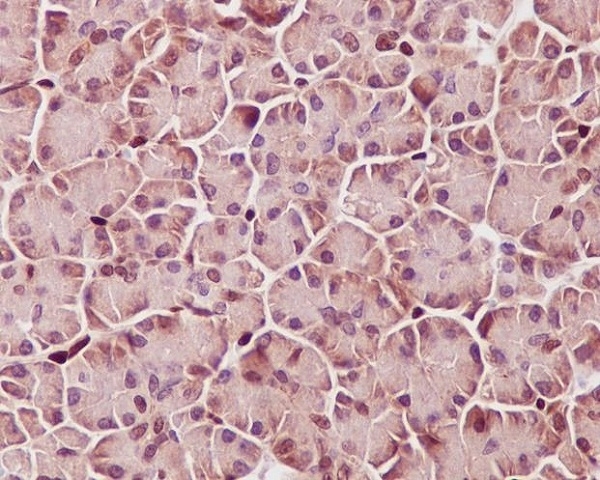
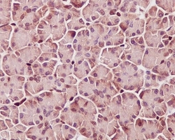
- Immunohistochemical analysis of paraffin-embedded human pancreas, using Phospho-STAT3 (Y705) Antibody(P00007-2)STAT3 was detected in paraffin-embedded tissue section. Heat mediated antigen retrieval was performed in citrate buffer (pH6, epitope retrieval solution) for 20 mins. The tissue section was blocked with 10% goat serum. The tissue section was then incubated with 1ug/ml rabbit anti-STAT3 Antibody (P00007-2)overnight at 4°C. Biotinylated goat anti-rabbit IgG was used as secondary antibody and incubated for 30 minutes at 37°C. The tissue section was developed using Strepavidin-Biotin-Complex (SABC)(Catalog # SA1022) with DAB as the chromogen.
- Additional image