 Explore
 Validate
 Learn
Learn Western blot
 Immunocytochemistry
Antibody data
- Antibody Data
- Antigen structure
- References [13]
- Comments [0]
- Validations
- Immunocytochemistry [4]
- Immunoprecipitation [1]
- Immunohistochemistry [1]
- Chromatin Immunoprecipitation [2]
- Other assay [8]
Submit
Validation data
Reference
Comment
Report error
- Product number
- MA1-13042 - Provider product page

- Provider
- Invitrogen Antibodies
- Product name
- STAT3 Monoclonal Antibody (9D8)
- Antibody type
- Monoclonal
- Antigen
- Recombinant full-length protein
- Description
- MA1-13042 detects STAT3 from human, mouse, monkey and rat samples. MA1-13042 has been successfully used in Western blot, ChIP, immunofluorescence, immunohistochemistry (paraffin), and immunoprecipitation procedures. By Western blot, this antibody detects a 88 kDa protein corresponding to STAT3. Positive control: HepG2 cells. The MA1-13042 antigen is a recombinant protein corresponding to residues 655-770 of human STAT3. Antibodies to this protein (and modification) were previously sold as part of a Thermo Scientific Cellomics High Content Screening Kit. This replacement antibody is now recommended for researchers who need an antibody for high content cell based assays. It has been thoroughly tested and validated for cellular immunofluorescence (IF) applications. Further optimization including the selection of the most appropriate fluorescent Dylight conjugated secondary antibody may have to be performed for your high content assay.
- Reactivity
- Human, Mouse, Rat
- Host
- Mouse
- Isotype
- IgG
- Antibody clone number
- 9D8
- Vial size
- 100 μL
- Concentration
- 1 mg/mL
- Storage
- -20°C, Avoid Freeze/Thaw Cycles
Submitted references Myelin repair is fostered by the corticosteroid medrysone specifically acting on astroglial subpopulations.
Abi1 mediates airway smooth muscle cell proliferation and airway remodeling via Jak2/STAT3 signaling.
Inhibition of IL-6 in the LCWE Mouse Model of Kawasaki Disease Inhibits Acute Phase Reactant Serum Amyloid A but Fails to Attenuate Vasculitis.
Discovery of a novel potentially transforming somatic mutation in CSF2RB gene in breast cancer.
miRNAs Potentially Involved in Post Lung Transplant-Obliterative Bronchiolitis: The Role of miR-21-5p.
The CDK4/6-EZH2 pathway is a potential therapeutic target for psoriasis.
MicroRNA-325-3p Facilitates Immune Escape of Mycobacterium tuberculosis through Targeting LNX1 via NEK6 Accumulation to Promote Anti-Apoptotic STAT3 Signaling.
Periostin Activation of Integrin Receptors on Sensory Neurons Induces Allergic Itch.
Fatty acids and cancer-amplified ZDHHC19 promote STAT3 activation through S-palmitoylation.
Effects of cell-cell crosstalk on gene expression patterns in a cell model of renal cell carcinoma lung metastasis.
An Autocrine Cytokine/JAK/STAT-Signaling Induces Kynurenine Synthesis in Multidrug Resistant Human Cancer Cells.
Whole-genome sequencing reveals oncogenic mutations in mycosis fungoides.
FGF2 regulates melanocytes viability through the STAT3-transactivated PAX3 transcription.
Silva Oliveira Junior M, Schira-Heinen J, Reiche L, Han S, de Amorim VCM, Lewen I, Gruchot J, Göttle P, Akkermann R, Azim K, Küry P
EBioMedicine 2022 Sep;83:104204
EBioMedicine 2022 Sep;83:104204
Abi1 mediates airway smooth muscle cell proliferation and airway remodeling via Jak2/STAT3 signaling.
Wang R, Wang Y, Liao G, Chen B, Panettieri RA Jr, Penn RB, Tang DD
iScience 2022 Feb 18;25(2):103833
iScience 2022 Feb 18;25(2):103833
Inhibition of IL-6 in the LCWE Mouse Model of Kawasaki Disease Inhibits Acute Phase Reactant Serum Amyloid A but Fails to Attenuate Vasculitis.
Porritt RA, Chase Huizar C, Dick EJ, Kumar S, Escalona R, Gomez AC, Marek-Iannucci S, Noval Rivas M, Patterson J, Forsthuber TG, Arditi M, Gorelik M
Frontiers in immunology 2021;12:630196
Frontiers in immunology 2021;12:630196
Discovery of a novel potentially transforming somatic mutation in CSF2RB gene in breast cancer.
Rashid M, Ali R, Almuzzaini B, Song H, AlHallaj A, Abdulkarim AA, Mohamed Baz O, Al Zahrani H, Mustafa Sabeena M, Alharbi W, Hussein M, Boudjelal M
Cancer medicine 2021 Nov;10(22):8138-8150
Cancer medicine 2021 Nov;10(22):8138-8150
miRNAs Potentially Involved in Post Lung Transplant-Obliterative Bronchiolitis: The Role of miR-21-5p.
Bozzini S, Pandolfi L, Rossi E, Inghilleri S, Zorzetto M, Ferrario G, Di Carlo S, Politano G, De Silvestri A, Frangipane V, Porzio M, Kessler R, Calabrese F, Meloni F, Morbini P
Cells 2021 Mar 20;10(3)
Cells 2021 Mar 20;10(3)
The CDK4/6-EZH2 pathway is a potential therapeutic target for psoriasis.
Müller A, Dickmanns A, Resch C, Schäkel K, Hailfinger S, Dobbelstein M, Schulze-Osthoff K, Kramer D
The Journal of clinical investigation 2020 Nov 2;130(11):5765-5781
The Journal of clinical investigation 2020 Nov 2;130(11):5765-5781
MicroRNA-325-3p Facilitates Immune Escape of Mycobacterium tuberculosis through Targeting LNX1 via NEK6 Accumulation to Promote Anti-Apoptotic STAT3 Signaling.
Fu B, Xue W, Zhang H, Zhang R, Feldman K, Zhao Q, Zhang S, Shi L, Pavani KC, Nian W, Lin X, Wu H
mBio 2020 Jun 2;11(3)
mBio 2020 Jun 2;11(3)
Periostin Activation of Integrin Receptors on Sensory Neurons Induces Allergic Itch.
Mishra SK, Wheeler JJ, Pitake S, Ding H, Jiang C, Fukuyama T, Paps JS, Ralph P, Coyne J, Parkington M, DeBrecht J, Ehrhardt-Humbert LC, Cruse GP, Bäumer W, Ji RR, Ko MC, Olivry T
Cell reports 2020 Apr 7;31(1):107472
Cell reports 2020 Apr 7;31(1):107472
Fatty acids and cancer-amplified ZDHHC19 promote STAT3 activation through S-palmitoylation.
Niu J, Sun Y, Chen B, Zheng B, Jarugumilli GK, Walker SR, Hata AN, Mino-Kenudson M, Frank DA, Wu X
Nature 2019 Sep;573(7772):139-143
Nature 2019 Sep;573(7772):139-143
Effects of cell-cell crosstalk on gene expression patterns in a cell model of renal cell carcinoma lung metastasis.
Kaminska K, Czarnecka AM, Khan MI, Fendler W, Klemba A, Krasowski P, Bartnik E, Szczylik C
International journal of oncology 2018 Mar;52(3):768-786
International journal of oncology 2018 Mar;52(3):768-786
An Autocrine Cytokine/JAK/STAT-Signaling Induces Kynurenine Synthesis in Multidrug Resistant Human Cancer Cells.
Campia I, Buondonno I, Castella B, Rolando B, Kopecka J, Gazzano E, Ghigo D, Riganti C
PloS one 2015;10(5):e0126159
PloS one 2015;10(5):e0126159
Whole-genome sequencing reveals oncogenic mutations in mycosis fungoides.
McGirt LY, Jia P, Baerenwald DA, Duszynski RJ, Dahlman KB, Zic JA, Zwerner JP, Hucks D, Dave U, Zhao Z, Eischen CM
Blood 2015 Jul 23;126(4):508-19
Blood 2015 Jul 23;126(4):508-19
FGF2 regulates melanocytes viability through the STAT3-transactivated PAX3 transcription.
Dong L, Li Y, Cao J, Liu F, Pier E, Chen J, Xu Z, Chen C, Wang RA, Cui R
Cell death and differentiation 2012 Apr;19(4):616-22
Cell death and differentiation 2012 Apr;19(4):616-22
No comments: Submit comment
Supportive validation
- Submitted by
- Invitrogen Antibodies (provider)
- Main image
- Experimental details
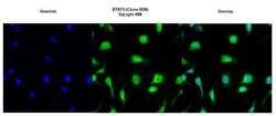
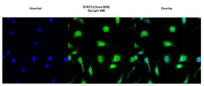
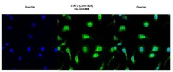
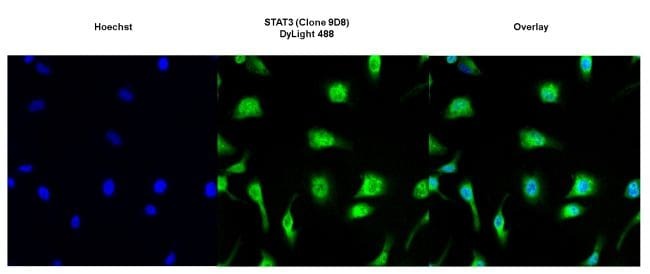
- Immunofluorescent analysis of STAT3 using anti-STAT3 (9D8) monoclonal antibody (Product # MA1-13042) (shown in green) in HeLa cells. Formalin fixed cells were permeabilized with 0.1% Triton X-100 in TBS for 10 minutes at room temperature. Cells were then blocked with 1% Blocker BSA (Product # 37525) for 15 minutes at room temperature. Cells were probed with a mouse monoclonal antibody recognizing STAT3 (Product # MA1-13042), at a dilution of 1:100 for at least 1 hour at room temperature. Cells were then washed with PBS and incubated with DyLight 488 goat-anti-mouse secondary antibody (Product # 35503) at a dilution of 1:400 for 30 minutes at room temperature. Nuclei (blue) were stained with Hoechst 33342 dye (Product # 62249). Images were taken on a Thermo Scientific ArrayScan at 20X magnification.
- Submitted by
- Invitrogen Antibodies (provider)
- Main image
- Experimental details
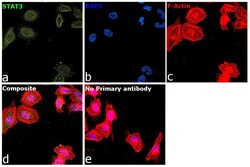
- Immunofluorescence analysis of Signal transducer and activator of transcription 3 was performed using 70% confluent log phase HeLa cells. The cells were fixed with 4% paraformaldehyde for 10 minutes, permeabilized with 0.1% Triton™ X-100 for 5 minutes, and blocked with 2% BSA for 45 minutes at room temperature. The cells were labeled with STAT3 Monoclonal Antibody (9D8) (Product # MA1-13042) at 1:100 dilution in 0.1% BSA, incubated at 4 degree celsius overnight and then labeled with Donkey anti-Mouse IgG (H+L) Highly Cross-Adsorbed Secondary Antibody, Alexa Fluor Plus 488 (Product # A32766), (1:2000 dilution), for 45 minutes at room temperature (Panel a: Green). Nuclei (Panel b:Blue) were stained with ProLong™ Diamond Antifade Mountant with DAPI (Product # P36962). F-actin (Panel c: Red) was stained with Rhodamine Phalloidin (Product # R415, 1:300). Panel d represents the merged image showing Nucleus and cytoplasm localization. Panel e represents control cells with no primary antibody to assess background. The images were captured at 60 magnification.

- Submitted by
- Invitrogen Antibodies (provider)
- Main image

- Experimental details
- Immunofluorescence analysis of Signal transducer and activator of transcription 3 was performed using 70% confluent log phase HeLa cells. The cells were fixed with 4% paraformaldehyde for 10 minutes, permeabilized with 0.1% Triton™ X-100 for 5 minutes, and blocked with 2% BSA for 45 minutes at room temperature. The cells were labeled with STAT3 Monoclonal Antibody (9D8) (Product # MA1-13042) at 1:100 dilution in 0.1% BSA, incubated at 4 degree celsius overnight and then labeled with Donkey anti-Mouse IgG (H+L) Highly Cross-Adsorbed Secondary Antibody, Alexa Fluor Plus 488 (Product # A32766), (1:2000 dilution), for 45 minutes at room temperature (Panel a: Green). Nuclei (Panel b:Blue) were stained with ProLong™ Diamond Antifade Mountant with DAPI (Product # P36962). F-actin (Panel c: Red) was stained with Rhodamine Phalloidin (Product # R415, 1:300). Panel d represents the merged image showing Nucleus and cytoplasm localization. Panel e represents control cells with no primary antibody to assess background. The images were captured at 60 magnification.

- Submitted by
- Invitrogen Antibodies (provider)
- Main image
- Experimental details
- Immunofluorescent analysis of STAT3 using anti-STAT3 (9D8) monoclonal antibody (Product # MA1-13042) (shown in green) in HeLa cells. Formalin fixed cells were permeabilized with 0.1% Triton X-100 in TBS for 10 minutes at room temperature. Cells were then blocked with 1% Blocker BSA (Product # 37525) for 15 minutes at room temperature. Cells were probed with a mouse monoclonal antibody recognizing STAT3 (Product # MA1-13042), at a dilution of 1:100 for at least 1 hour at room temperature. Cells were then washed with PBS and incubated with DyLight 488 goat-anti-mouse secondary antibody (Product # 35503) at a dilution of 1:400 for 30 minutes at room temperature. Nuclei (blue) were stained with Hoechst 33342 dye (Product # 62249). Images were taken on a Thermo Scientific ArrayScan at 20X magnification.
Supportive validation
- Submitted by
- Invitrogen Antibodies (provider)
- Main image
- Experimental details
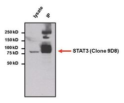
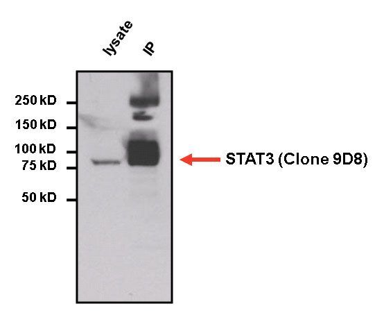
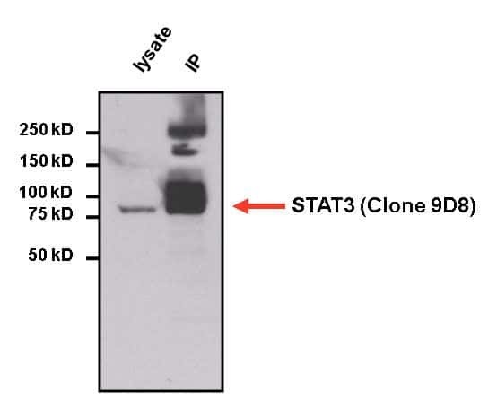
- Immunoprecipitation of STAT3 was performed on HepG2 cells. The antigen:antibody complex was formed by incubating 750 µg whole cell lysate with 2 µg of mouse monoclonal antibody recognizing STAT3 (Product # MA1-13042) overnight on a rocking platform at 4°C. The immune-complex was then captured on 50 µL Protein A/G Plus Agarose (Product # 20423). Captured immune-complexes were then washed extensively and proteins eluted with 5X Reducing Sample Loading Dye (Product # 39000). Samples were then resolved on a 4-20% Tris-HCl polyacrylamide gel. Proteins were transferred to PVDF membrane and blocked with 5% Milk/TBS-0.1%Tween for at least 1 hour. Membranes were then probed with a mouse monoclonal antibody recognizing STAT3 (Product # MA1-13042) at a dilution of 1:5000 overnight rotating at 4°C. Membranes were washed in TBST and probed with Clean-blot IP detection reagent (Product # 21230) at a dilution of 1:2000 for at least one hour. Membranes were washed and chemiluminescent detection was performed using Pierce Super Signal West Dura (Product # 34075).
Supportive validation
- Submitted by
- Invitrogen Antibodies (provider)
- Main image
- Experimental details
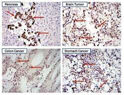
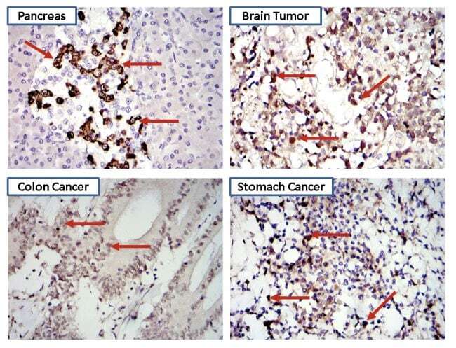
- Immunohistochemistry was performed on biopsies of normal and cancer tissues. To expose target proteins heat induced antigen retrieval was performed using 10mM sodium citrate (pH6.0) buffer for 20 minutes at 95°C. Following antigen retrieval tissues were blocked in 10% normal goat serum (Product # 31873) for 20 minutes at room temperature. Tissues were then probed at a dilution of 1:1600 with a mouse monoclonal antibody recognizing STAT3 (Product # MA1-13042) overnight at 4°C in a humidified chamber. Detection was performed using a goat anti-mouse HRP secondary antibody followed by colorimetric detection using DAB substrate. Tissues were counterstained with hematoxylin and prepped for mounting. Images were taken at 400X magnification. Results demonstrate both nuclear and cytoplasmic localization of STAT3.
Supportive validation
- Submitted by
- Invitrogen Antibodies (provider)
- Main image
- Experimental details
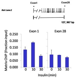
- Chromatin immunoprecipitation analysis of STAT3 was performed using cross-linked chromatin from 1x10^6 HTC-IR rat hepatoma cells treated with insulin for 0, 10, and 30 minutes. Immunoprecipitation was performed using a multiplex microplate Matrix ChIP assay (see reference for Matrix ChIP protocol: http://www.ncbi.nlm.nih.gov/pubmed/22098709) with 1.0 µL/100 µL well volume of a STAT3 monoclonal antibody (Product # MA1-13042). Chromatin aliquots from ~1x10^5 cells were used per ChIP pull-down. Quantitative PCR data were done in quadruplicate using 1 µL of eluted DNA in 2 µL SYBR real-time PCR reactions containing primers to amplify exon-1 or exon-28 of the LAMC1 gene. PCR calibration curves were generated for each primer pair from a dilution series of sheared total genomic DNA. Quantitation of immunoprecipitated chromatin is presented as signal relative to the total amount of input chromatin. Results represent the mean +/- SEM for three experiments. A schematic representation of the rat LAMC1 locus is shown above the data where boxes represent exons (black boxes = translated regions, white boxes = untranslated regions), the zigzag line represents an intron, and the straight line represents upstream sequence. Regions amplified by LAMC1 primers are represented by black bars. Data courtesy of the Innovators Program.
- Submitted by
- Invitrogen Antibodies (provider)
- Main image

- Experimental details
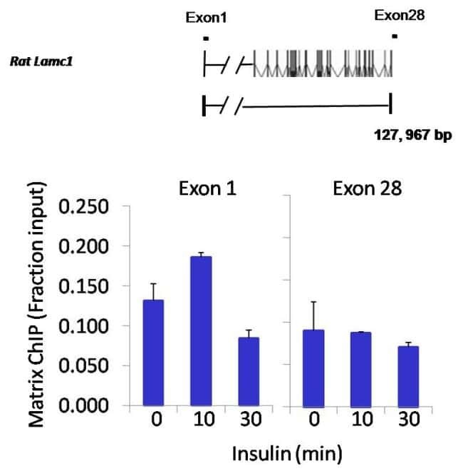
- Chromatin immunoprecipitation analysis of STAT3 was performed using cross-linked chromatin from 1x10^6 HTC-IR rat hepatoma cells treated with insulin for 0, 10, and 30 minutes. Immunoprecipitation was performed using a multiplex microplate Matrix ChIP assay (see reference for Matrix ChIP protocol: http://www.ncbi.nlm.nih.gov/pubmed/22098709) with 1.0 µL/100 µL well volume of a STAT3 monoclonal antibody (Product # MA1-13042). Chromatin aliquots from ~1x10^5 cells were used per ChIP pull-down. Quantitative PCR data were done in quadruplicate using 1 µL of eluted DNA in 2 µL SYBR real-time PCR reactions containing primers to amplify exon-1 or exon-28 of the LAMC1 gene. PCR calibration curves were generated for each primer pair from a dilution series of sheared total genomic DNA. Quantitation of immunoprecipitated chromatin is presented as signal relative to the total amount of input chromatin. Results represent the mean +/- SEM for three experiments. A schematic representation of the rat LAMC1 locus is shown above the data where boxes represent exons (black boxes = translated regions, white boxes = untranslated regions), the zigzag line represents an intron, and the straight line represents upstream sequence. Regions amplified by LAMC1 primers are represented by black bars. Data courtesy of the Innovators Program.

Supportive validation
- Submitted by
- Invitrogen Antibodies (provider)
- Main image

- Experimental details
- Immunoprecipitation of STAT3 was performed on HepG2 cells. The antigen:antibody complex was formed by incubating 750 µg whole cell lysate with 2 µg of mouse monoclonal antibody recognizing STAT3 (Product # MA1-13042) overnight on a rocking platform at 4øC. The immune-complex was then captured on 50 µL Protein A/G Plus Agarose (Product # 20423). Captured immune-complexes were then washed extensively and proteins eluted with 5X Reducing Sample Loading Dye (Product # 39000). Samples were then resolved on a 4-20% Tris-HCl polyacrylamide gel. Proteins were transferred to PVDF membrane and blocked with 5% Milk/TBS-0.1%Tween for at least 1 hour. Membranes were then probed with a mouse monoclonal antibody recognizing STAT3 (Product # MA1-13042) at a dilution of 1:5000 overnight rotating at 4øC. Membranes were washed in TBST and probed with Clean-blot IP detection reagent (Product # 21230) at a dilution of 1:2000 for at least one hour. Membranes were washed and chemiluminescent detection was performed using Pierce Super Signal West Dura (Product # 34075).
- Submitted by
- Invitrogen Antibodies (provider)
- Main image
- Experimental details
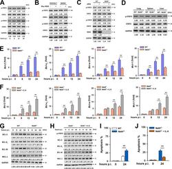
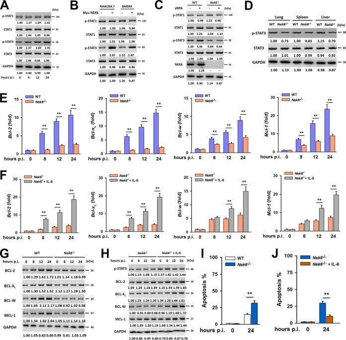
- FIG 5 NEK6 inhibits apoptosis through the activation of STAT3. (A) Immunoblot analysis of the phosphorylated STAT1 (p-STAT1), STAT1, phosphorylated STAT3 (p-STAT3), and STAT3 in RAW 264.7 cells stimulated with 10 mug/mL of gamma-irradiated M. tuberculosis . (B) Immunoblot analysis of the p-STAT1, STAT1, p-STAT3, and STAT3 in RAW 264.7 cells and BMDMs transfected with Myc-NEK6. (C) BMDMs from WT and NEK6-deficient ( Nek6 -/- ) mice were stimulated with 10 mug/mL of gamma-irradiated M. tuberculosis for 24 h. The expressions of p-STAT1, STAT1, p-STAT3, STAT3, and NEK6 were analyzed by Western blotting. (D) Immunoblot analysis of the p-STAT3 and STAT3 in lungs, spleens, and livers from WT and Nek6 -/- mice at 7 dpi. (E and G) The expression levels of BCL-2, BCL-X L , BCL-W, and MCL-1 in 10 mug/mL of gamma-irradiated M. tuberculosis -stimulated BMDMs from WT and Nek6 -/- mice were calculated by qRT-PCR at the indicated times (E) and by Western blotting (G). (F and H) BMDMs from Nek6 -/- mice were pretreated with 20 ng/mL IL-6 for 4 h h, then cells were stimulated with 10 mug/mL of gamma-irradiated M. tuberculosis . The expression levels of BCL-2, BCL-X L , BCL-W, and MCL-1 were calculated by qRT-PCR (F) and Western blotting (H) at the indicated times. (I) BMDMs from WT and Nek6 -/- mice were stimulated with 10 mug/mL of gamma-irradiated M. tuberculosis for 24 h, and the apoptosis rates were detected by flow cytometry. (J) BMDMs from Nek6 -/- mice were pretreated with 20 ng/mL of I
- Submitted by
- Invitrogen Antibodies (provider)
- Main image
- Experimental details
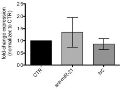
- Figure 6 Western blot of STAT3 protein expression levels in LFs treated with anti-miR-21. Quantification of immunoblots using anti-STAT3 of LFs from BAL of CLAD patients treated with anti-miR-21 after 48h. Control cells (CTR); negative control treated cells (NC).

- Submitted by
- Invitrogen Antibodies (provider)
- Main image
- Experimental details
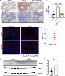
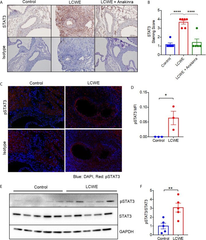
- Figure 2 STAT3 expression in the LCWE-induced model of vasculitis and response to anakinra treatment. (A) Representative images of immunohistochemistry staining for STAT3 in murine heart of PBS control, LCWE injected or LCWE injected mice treated with anakinra. (B) STAT3 staining score in PBS control, LCWE injected and LCWE injected mice treated with anakinra. (C) Representative images of pSTAT3 immunofluorescence staining in murine heart tissue from PBS control and LCWE injected mice. (D) Quantification of pSTAT3 immunofluorescence staining in murine heart tissue. (E) Western blot of pSTAT3 and STAT3 in murine heart tissue from PBS control and LCWE injected mice. (F) Quantification of pSTAT3/STAT3 ratio from Western Blot. Data was analyzed by one-way ANOVA with Tukey''s multiple testing comparison (B) or Student''s t test (D, F) . * p < 0.05, ** p < 0.01, **** p < 0.0001.
- Submitted by
- Invitrogen Antibodies (provider)
- Main image
- Experimental details
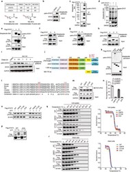
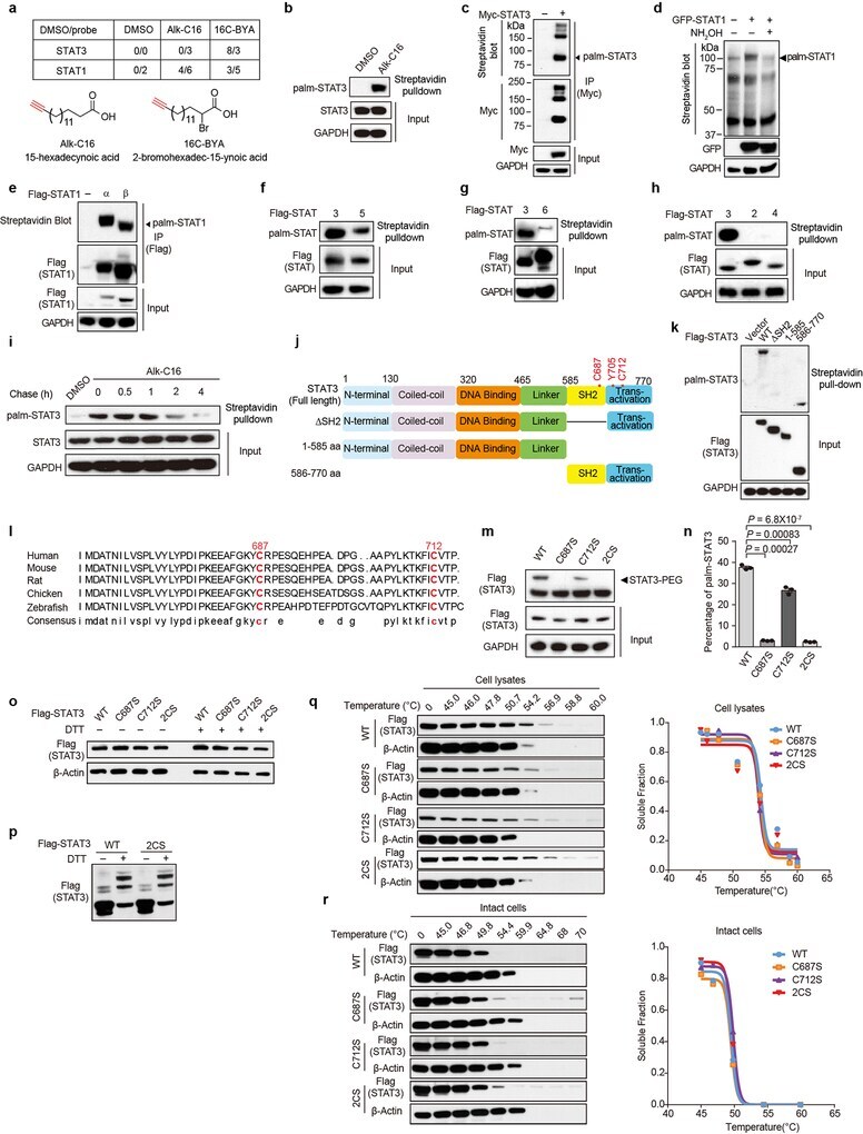
- Extended Data Figure 1. STAT3 is S-palmitoylated at evolutionarily conserved cysteine residues and mutation of STAT3 palmitoylation sites does not affect disulfide formation and protein stability. (a) HEK293A cells were incubated for 4 hours with DMSO or 50 muM clickable probes. Cell lysates were reacted with azide-biotin for enrichment of labeled proteins with streptavidin beads and identified by mass spectrometry. The peptide spectral counts were shown in the table. ( b ) HEK293A cells were labeled with 50 muM chemical probe (Alk-C16) for 4 hours. Cell lysates were reacted with biotin-azide and precipitated with streptavidin beads and subjected to western blot using anti-STAT3 antibody. Endogenous STAT3 palmitoylation was analyzed by western blot. (c) HEK293A cells were transfected with Myc-STAT3 WT and labeled with 50 muM chemical probe Alk-C16 for 4 hours, cell lysates were subjected to Streptavidin blot following anti-Myc IP and subsequent Click reaction. (d) HEK293A cells were transfected with empty vector or GFP-STAT1 and labeled with 50 muM chemical probe Alk-C16 for 4 hours. After Click reaction, cell lysates were subjected to streptavidin blot showing detection of STAT1. (e) Similar analysis as panel (c) was performed in HEK293A cells transfected with Flag-STAT1alpha and Flag-STAT1beta. (f) HEK293A cells were transfected with Flag-STAT3 or Flag-STAT5 and labeled with 50 muM chemical probe Alk-C16 for 4 hours, Cell lysates were reacted with biotin-azide and precipita
- Submitted by
- Invitrogen Antibodies (provider)
- Main image
- Experimental details
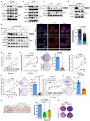
- Extended Data Figure 7. ZDHHC19 mediated STAT3 palmitoylaiton facilitates LSCC tumor cell growth, colony formation and migration in vitro through induction of STAT3 activity. (a) HCC95 lung squamous cell carcinoma cells were pretreated with ruxolitinib (1 muM) for 30 minutes and then labeled with 50 muM probe Alk-C16 for 2 hours with the incubation of IL-6 (20 ng/ml). Endogenous STAT3 palmitoylation was analyzed by Click reaction and streptavidin beads pulldown, and followed by western blotting. (b) HCC95 cells were treated as in panel (a). Whole cell lysates were analyzed by anti-STAT3 IP followed by western blotting using the indicated antibodies. (c) Lung squamous cell carcinoma cell lines HCC95 and KNS-62 were transfected with shRNA control or shZDHHC19 (3 and 5) and labeled with 50 muM Alk-C16 probe for 4 hours, Cell lysates were reacted with biotin-azide and precipitated with streptavidin beads. Endogenous STAT3 palmitoylation was determined by western blot. (d) SK-MES-1 cells were transfected with ZDHHC19-shRNA and labeled with 50 muM palmitoylation probe Alk-C16 for 4hours. Cells lysates were reacted with azide-biotin and subjected to streptavidin beads pull-down. STAT3 palmitoylation was shown by western blot. (e) HCC95 shRNA control cells and Sh3 ZDHHC19 stable cells were pretreated with ruxolitinib (1 muM) for 30 minutes and then labeled with 50 muM probe Alk-C16 for 2 hours with the incubation of IL-6 (20 ng/ml). Endogenous STAT3 palmitoylation was determined by w

- Submitted by
- Invitrogen Antibodies (provider)
- Main image
- Experimental details
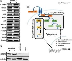
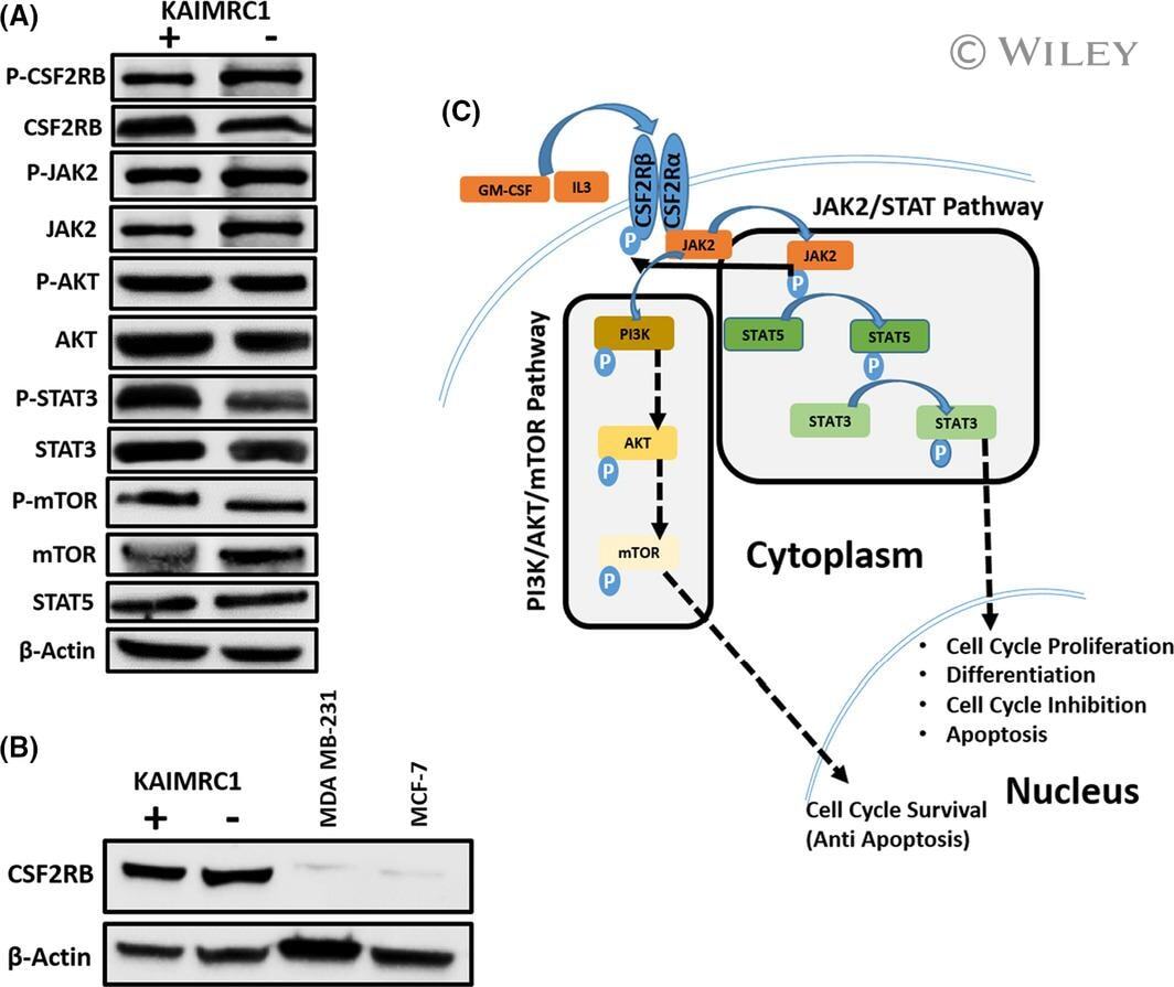
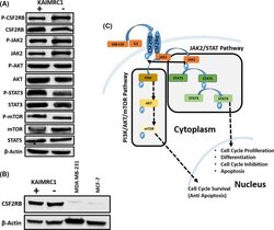
- 2 FIGURE Characterization of KAIMRC1 cells on protein level. (A) Western blot analysis of the KAIMRC1 cell line in normal (+) and serum-starved conditions(-) against P- CSF2RB , CSF2RB , P-AKT1, AKT1, JAK2, P-JAK2, STAT3, P-STAT3, STAT5, mTOR, and P-mTOR. Ligand-independent activation of AKT/mTOR pathway, as well as the JAK2/STAT pathway, was observed. (B) Comparative Western blot analysis of KAIMRC1, MDA-MB-231, and MCF-7 cell lines against CSF2RB . (C) Schematic representation of proposed activation of the AKT/mTOR pathway and JAK2/STAT pathway that might result in cell cycle survival and proliferation
- Submitted by
- Invitrogen Antibodies (provider)
- Main image
- Experimental details
- FIGURE 2 Characterization of KAIMRC1 cells on protein level. (A) Western blot analysis of the KAIMRC1 cell line in normal (+) and serum-starved conditions(-) against P- CSF2RB , CSF2RB , P-AKT1, AKT1, JAK2, P-JAK2, STAT3, P-STAT3, STAT5, mTOR, and P-mTOR. Ligand-independent activation of AKT/mTOR pathway, as well as the JAK2/STAT pathway, was observed. (B) Comparative Western blot analysis of KAIMRC1, MDA-MB-231, and MCF-7 cell lines against CSF2RB . (C) Schematic representation of proposed activation of the AKT/mTOR pathway and JAK2/STAT pathway that might result in cell cycle survival and proliferation
