 Explore
 Validate
 Learn
Learn Western blot
Antibody data
- Antibody Data
- Antigen structure
- References [2]
- Comments [0]
- Validations
- Western blot [1]
- Immunocytochemistry [1]
- Flow cytometry [1]
Submit
Validation data
Reference
Comment
Report error
- Product number
- AF4607 - Provider product page

- Provider
- R&D Systems
- Product name
- Human Phospho-STAT3 (Y705) Antibody
- Antibody type
- Polyclonal
- Description
- Antigen Affinity-purified. Detects human STAT3 when phosphorylated at Y705.
- Reactivity
- Human
- Host
- Rabbit
- Conjugate
- Unconjugated
- Isotype
- IgG
- Vial size
- 100 ug
- Concentration
- LYOPH
- Storage
- Use a manual defrost freezer and avoid repeated freeze-thaw cycles. 12 months from date of receipt, -20 to -70 °C as supplied. 1 month, 2 to 8 °C under sterile conditions after reconstitution. 6 months, -20 to -70 °C under sterile conditions after reconstitution.
Submitted references Extracellular vesicles secreted by hypoxia pre-challenged mesenchymal stem cells promote non-small cell lung cancer cell growth and mobility as well as macrophage M2 polarization via miR-21-5p delivery.
IL-6 cytoprotection in hyperoxic acute lung injury occurs via suppressor of cytokine signaling-1-induced apoptosis signal-regulating kinase-1 degradation.
Ren W, Hou J, Yang C, Wang H, Wu S, Wu Y, Zhao X, Lu C
Journal of experimental & clinical cancer research : CR 2019 Feb 8;38(1):62
Journal of experimental & clinical cancer research : CR 2019 Feb 8;38(1):62
IL-6 cytoprotection in hyperoxic acute lung injury occurs via suppressor of cytokine signaling-1-induced apoptosis signal-regulating kinase-1 degradation.
Kolliputi N, Waxman AB
American journal of respiratory cell and molecular biology 2009 Mar;40(3):314-24
American journal of respiratory cell and molecular biology 2009 Mar;40(3):314-24
No comments: Submit comment
Supportive validation
- Submitted by
- R&D Systems (provider)
- Main image

- Experimental details
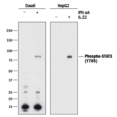
- Detection of Human Phospho-STAT3 (Y705) by Western Blot. Western blot shows lysates of Daudi human Burkitt's lymphoma cell line and HepG2 human hepatocellular carcinoma cell line untreated (-) or treated (+) with 500 U/mL Recombinant Human IFN-alpha A (Catalog # 11100-1) for 20 minutes or 50 μg/mL Recombinant Human IL-22 (Catalog # 782-IL) for 15 minutes. PVDF membrane was probed with 0.5 µg/mL of Rabbit Anti-Human Phospho-STAT3 (Y705) Antigen Affinity-purified Polyclonal Antibody (Catalog # AF4607) followed by HRP-conjugated Anti-Rabbit IgG Secondary Antibody (Catalog # HAF008). A specific band was detected for STAT3 at approximately 95 kDa (as indicated). This experiment was conducted under reducing conditions and using Immunoblot Buffer Group 1.
Supportive validation
- Submitted by
- R&D Systems (provider)
- Main image
- Experimental details
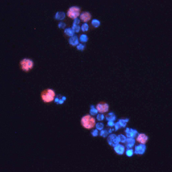
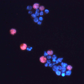
- STAT3 in Daudi Human Burkitt's Lymphoma Cells. Phospho-STAT3 was detected in immersion fixed IFN-alpha treated Daudi human Burkitt's lymphoma cell line using 10 µg/mL Human Phospho-STAT3 (Y705) Antigen Affinity-purified Polyclonal Antibody (Catalog # AF4607) for 3 hours at room temperature. Cells were stained with the NorthernLights™ 557-conjugated Anti-Rabbit IgG Secondary Antibody (red; Catalog # NL004) and counterstained with DAPI (blue). View our protocol for Fluorescent ICC Staining of Cells on Coverslips.
Supportive validation
- Submitted by
- R&D Systems (provider)
- Main image
- Experimental details
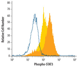
- Detection of Phospho-STAT3 (Y705) in IFN-alpha-treated Human Daudi Cell Line by Flow Cytometry. Daudi human Burkitt's lymphoma cell line was unstimulated (light orange filled histogram) or treated with 500 U/mL rhIFN-alpha for 20 minutes (dark orange filled histogram) was stained with Human Phospho-STAT3 (Y705) Antigen Affinity-purified Polyclonal Antibody (Catalog # AF4607) or control antibody (Catalog # AB-105-C, open histogram), followed by Phycoerythrin-conjugated Anti-Rabbit IgG Secondary Antibody (Catalog # F0110). To facilitate intracellular staining, cells were fixed with paraformaldehyde and permeabilized with methanol.
