 Explore
 Validate
 Learn
Learn Western blot
 Immunoprecipitation
Antibody data
- Antibody Data
- Antigen structure
- References [5]
- Comments [0]
- Validations
- Western blot [2]
- Immunocytochemistry [2]
- Flow cytometry [2]
Submit
Validation data
Reference
Comment
Report error
- Product number
- MAB1799 - Provider product page

- Provider
- R&D Systems
- Product name
- Human/Mouse/Rat STAT3 Antibody
- Antibody type
- Monoclonal
- Description
- Protein A or G purified from hybridoma culture supernatant. Detects human, mouse, and rat STAT3 in Western blots.
- Reactivity
- Human, Mouse, Rat
- Host
- Mouse
- Conjugate
- Unconjugated
- Antigen sequence
P40763- Isotype
- IgG
- Antibody clone number
- 232209
- Vial size
- 100 ug
- Storage
- Use a manual defrost freezer and avoid repeated freeze-thaw cycles. 12 months from date of receipt, -20 to -70 °C, as supplied. 1 month, 2 to 8 °C under sterile conditions after opening. 6 months, -20 to -70 °C under sterile conditions after opening.
Submitted references Opa interacting protein 5 promotes metastasis of nasopharyngeal carcinoma cells by promoting EMT via modulation of JAK2/STAT3 signal.
Tumor-derived mesenchymal-stem-cell-secreted IL-6 enhances resistance to cisplatin via the STAT3 pathway in breast cancer.
Inhibition of STAT3 Signaling Reduces IgA1 Autoantigen Production in IgA Nephropathy.
Proangiogenic tumor proteins as potential predictive or prognostic biomarkers for bevacizumab therapy in metastatic colorectal cancer.
IL-6 cytoprotection in hyperoxic acute lung injury occurs via suppressor of cytokine signaling-1-induced apoptosis signal-regulating kinase-1 degradation.
Zheng YQ, Cui YR, Yang S, Wang YP, Qiu YJ, Hu WL
European review for medical and pharmacological sciences 2019 Jan;23(2):613-621
European review for medical and pharmacological sciences 2019 Jan;23(2):613-621
Tumor-derived mesenchymal-stem-cell-secreted IL-6 enhances resistance to cisplatin via the STAT3 pathway in breast cancer.
Xu H, Zhou Y, Li W, Zhang B, Zhang H, Zhao S, Zheng P, Wu H, Yang J
Oncology letters 2018 Jun;15(6):9142-9150
Oncology letters 2018 Jun;15(6):9142-9150
Inhibition of STAT3 Signaling Reduces IgA1 Autoantigen Production in IgA Nephropathy.
Yamada K, Huang ZQ, Raska M, Reily C, Anderson JC, Suzuki H, Ueda H, Moldoveanu Z, Kiryluk K, Suzuki Y, Wyatt RJ, Tomino Y, Gharavi AG, Weinmann A, Julian BA, Willey CD, Novak J
Kidney international reports 2017 Nov;2(6):1194-1207
Kidney international reports 2017 Nov;2(6):1194-1207
Proangiogenic tumor proteins as potential predictive or prognostic biomarkers for bevacizumab therapy in metastatic colorectal cancer.
Bruhn MA, Townsend AR, Khoon Lee C, Shivasami A, Price TJ, Wrin J, Arentz G, Tebbutt NC, Hocking C, Cunningham D, Hardingham JE, BHI in collaboration with AGITG.
International journal of cancer 2014 Aug 1;135(3):731-41
International journal of cancer 2014 Aug 1;135(3):731-41
IL-6 cytoprotection in hyperoxic acute lung injury occurs via suppressor of cytokine signaling-1-induced apoptosis signal-regulating kinase-1 degradation.
Kolliputi N, Waxman AB
American journal of respiratory cell and molecular biology 2009 Mar;40(3):314-24
American journal of respiratory cell and molecular biology 2009 Mar;40(3):314-24
No comments: Submit comment
Supportive validation
- Submitted by
- R&D Systems (provider)
- Main image
- Experimental details
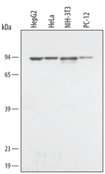
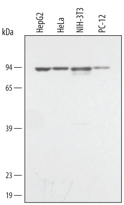
- Detection of Human STAT3 by Western Blot. Western blot shows lysates of HepG2 human hepatocellular carcinoma cell line, HeLa human cervical epithelial carcinoma cell line, NIH-3T3 mouse embryonic fibroblast cell line, and PC-12 rat adrenal pheochromocytoma cell line. PVDF membrane was probed with 0.1 µg/mL of Mouse Anti-Human STAT3 Monoclonal Antibody (Catalog # MAB1799) followed by HRP-conjugated Anti-Mouse IgG Secondary Antibody (Catalog # HAF007). A specific band was detected for STAT3 at approximately 90 kDa (as indicated). This experiment was conducted under reducing conditions and using Immunoblot Buffer Group 1.
- Submitted by
- R&D Systems (provider)
- Main image
- Experimental details
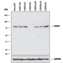
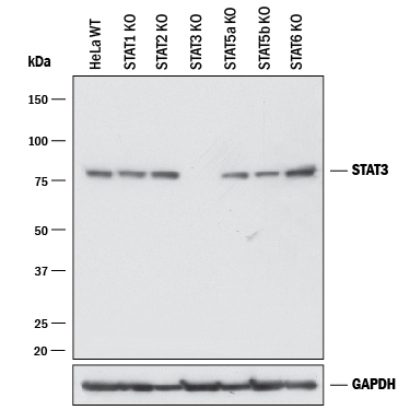
- Western Blot Shows Human STAT3 Specificity by Using Knockout Cell Line. Western blot shows lysates of HeLa human cervical epithelial carcinoma parental cell line, STAT1 knockout (KO) HeLa cell line, STAT2 KO HeLa cell line, STAT3 KO HeLa cell line, STAT5a KO HeLa cell line, STAT5b KO HeLa cell line, STAT6 KO HeLa cell line,. PVDF membrane was probed with 0.5 µg/mL of Mouse Anti-Human/Mouse/Rat STAT3 Monoclonal Antibody (Catalog # MAB1799) followed by HRP-conjugated Anti-Mouse IgG Secondary Antibody (Catalog # HAF018). A specific band was detected for STAT3 at approximately 80 kDa (as indicated) in the parental HeLa cell line, but is not detectable in the STAT3 knockout HeLa cell line. GAPDH (Catalog # AF5718) is shown as a loading control. This experiment was conducted under reducing conditions and using Immunoblot Buffer Group 1.
Supportive validation
- Submitted by
- R&D Systems (provider)
- Main image
- Experimental details
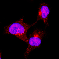
- STAT3 in HeLa Human Cell Line. STAT3 was detected in immersion fixed HeLa human cervical epithelial carcinoma cell line using Mouse Anti-Human/Mouse/Rat STAT3 Monoclonal Antibody (Catalog # MAB1799) at 3 µg/mL for 3 hours at room temperature. Cells were stained using the NorthernLights™ 557-conjugated Anti-Mouse IgG Secondary Antibody (red; Catalog # NL007) and counterstained with DAPI (blue). Specific staining was localized to cytoplasm and nuclei. View our protocol for Fluorescent ICC Staining of Cells on Coverslips.

- Submitted by
- R&D Systems (provider)
- Main image
- Experimental details
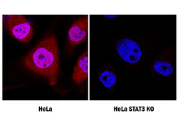
- STAT3 Specificity is Shown by Immunocytochemistry in Knockout Cell Line. STAT3 was detected in immersion fixed HeLa human cervical epithelial carcinoma cell line treated with IFN-alpha 1, but is not detected in STAT3 knockout (KO) HeLa cell line using Mouse Anti-Human/ Mouse/Rat STAT3 Monoclonal Antibody (Catalog # MAB1799) at 1 µg/mL for 3 hours at room temperature. Cells were stained using the NorthernLights 557-conjugated Anti-Mouse IgG Secondary Antibody (red; Catalog # NL007) and counterstained with DAPI (blue). Specific staining was localized to cytoplasm and nuclei. View our protocol for Fluorescent ICC Staining of Cells on Coverslips.

Supportive validation
- Submitted by
- R&D Systems (provider)
- Main image
- Experimental details
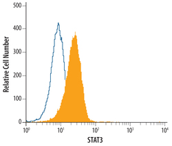
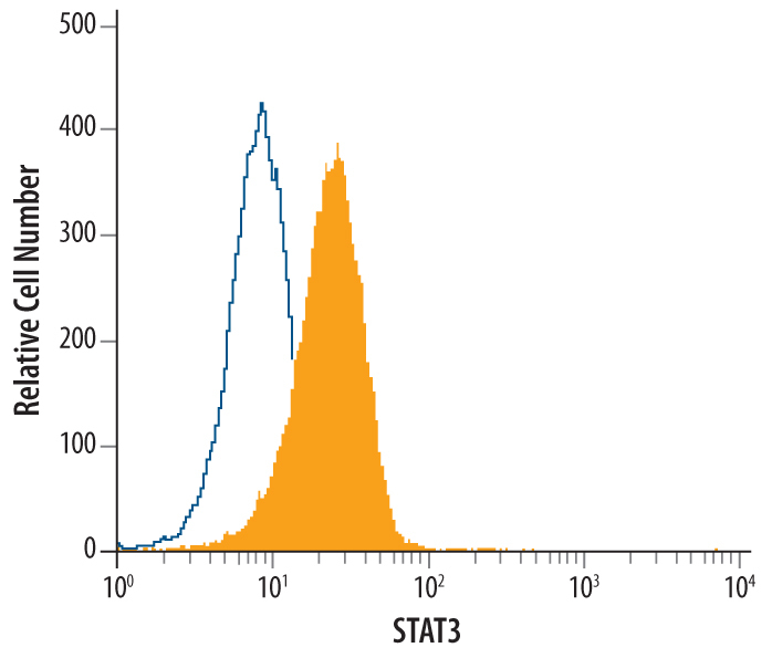
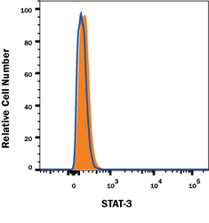
- Detection of STAT3 in Jurkat Human Cell Line by Flow Cytometry. Jurkat human acute T cell leukemia cell line was stained with Mouse Anti-Human STAT3 Monoclonal Antibody (Catalog # MAB1799, filled histogram) or isotype control antibody (Catalog # MAB0041, open histogram), followed by Allophycocyanin-conjugated Anti-Mouse IgG F(ab')2 Secondary Antibody (Catalog # F0101B). To facilitate intracellular staining, cells were fixed with paraformaldehyde and permeabilized with methanol.
- Submitted by
- R&D Systems (provider)
- Main image
- Experimental details
- STAT3 Specificity is Shown by Flow Cytometry in Knockout Cell Line. STAT3 knockout HeLa human cervical epithelial cell line was stained with Mouse Anti-Human/Mouse STAT3 Monoclonal Antibody (Catalog # MAB1799, filled histogram) or isotype control antibody (Catalog # MAB0041, open histogram) followed by anti-Mouse IgG PE-conjugated secondary antibody (Catalog # F0102B). No staining in the STAT3 knockout HeLa cell line was observed. To facilitate intracellular staining, cells were fixed with Flow Cytometry Fixation Buffer (Catalog # FC004) and permeabilized with Flow Cytometry Permeabilization/Wash Buffer I (Catalog # FC005). View our protocol for Staining Intracellular Molecules.
