Explore
 Validate
 Learn
Learn Western blot
 ELISA
ELISAAntibody data
- Antibody Data
- Antigen structure
- References [1]
- Comments [0]
- Validations
- Western blot [2]
- Immunocytochemistry [5]
- Chromatin Immunoprecipitation [2]
- Other assay [1]
Submit
Validation data
Reference
Comment
Report error
- Product number
- MA5-15659 - Provider product page

- Provider
- Invitrogen Antibodies
- Product name
- STAT6 Monoclonal Antibody (7D3)
- Antibody type
- Monoclonal
- Antigen
- Purifed from natural sources
- Description
- MA5-15659 targets STAT6 in indirect ELISA, IF and WB applications and shows reactivity with Human, mouse, Non-human primate, and Rat samples. The MA5-15659 immunogen is purified recombinant fragment of human STAT6 expressed in E. Coli. MA5-15659 detects STAT6 which has a predicted molecular weight of approximately 94kDa.
- Reactivity
- Human, Mouse, Rat
- Host
- Mouse
- Isotype
- IgG
- Antibody clone number
- 7D3
- Vial size
- 100 μL
- Concentration
- Conc. Not Determined
- Storage
- Store at 4°C short term. For long term storage, store at -20°C, avoiding freeze/thaw cycles.
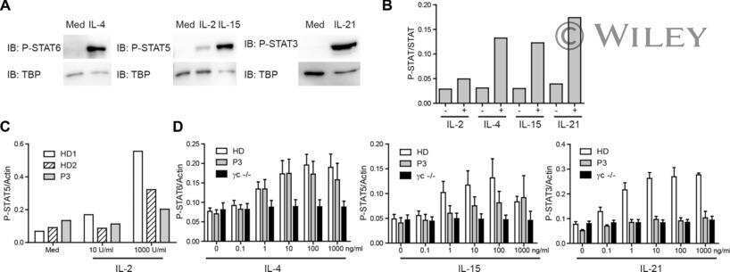
Submitted references Patients with T⁺/low NK⁺ IL-2 receptor γ chain deficiency have differentially-impaired cytokine signaling resulting in severe combined immunodeficiency.
Fuchs S, Rensing-Ehl A, Erlacher M, Vraetz T, Hartjes L, Janda A, Rizzi M, Lorenz MR, Gilmour K, de Saint-Basile G, Roifman CM, Cheuk S, Gennery A, Thrasher AJ, Fuchs I, Schwarz K, Speckmann C, Ehl S
European journal of immunology 2014 Oct;44(10):3129-40
European journal of immunology 2014 Oct;44(10):3129-40
No comments: Submit comment
Supportive validation
- Submitted by
- Invitrogen Antibodies (provider)
- Main image
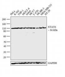
- Experimental details
- Western blot analysis was performed on whole cell extracts (30 µg lysate) of HeLa (Lane 1), K-562 (Lane 2), THP-1 (Lane 3), MOLT-4 (Lane 4), Jurkat (Lane 5), MCF7 (Lane 6), T-47D (Lane 7), SK-BR-3 (Lane 8), MDA-MB-231 (Lane 9) and HT-29 (Lane 10). The blot was probed with Anti-STAT6 Monoclonal Antibody (Product # MA5-15659, 1:500 dilution) and detected by chemiluminescence using Goat anti-Mouse IgG (H+L) Superclonal™ Secondary Antibody, HRP conjugate (Product # A28177, 0.25 µg/mL, 1:4000 dilution). A 94 kDa band corresponding to STAT6 was observed across the cell lines tested.
- Submitted by
- Invitrogen Antibodies (provider)
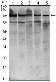
- Main image

- Experimental details
- Western blot analysis of STAT6 using STAT6 monoclonal antibody (Product # MA5-15659) in HEK293 (1), NIH/3T3 (2), MCF-7 (3), Raw246.7 (4) and PC-12 (5) cell lysate.
Supportive validation
- Submitted by
- Invitrogen Antibodies (provider)
- Main image

- Experimental details
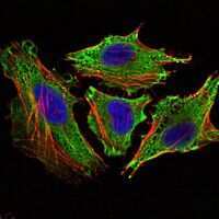
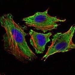
- Immunofluorescence analysis of HeLa cells using STAT6 monoclonal antibody (Product # MA5-15659) (Green). Blue: DRAQ5 fluorescent DNA dye. Red: actin filaments have been labeled with phalloidin.
- Submitted by
- Invitrogen Antibodies (provider)
- Main image
- Experimental details
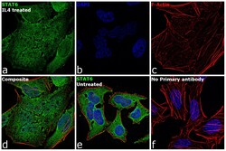
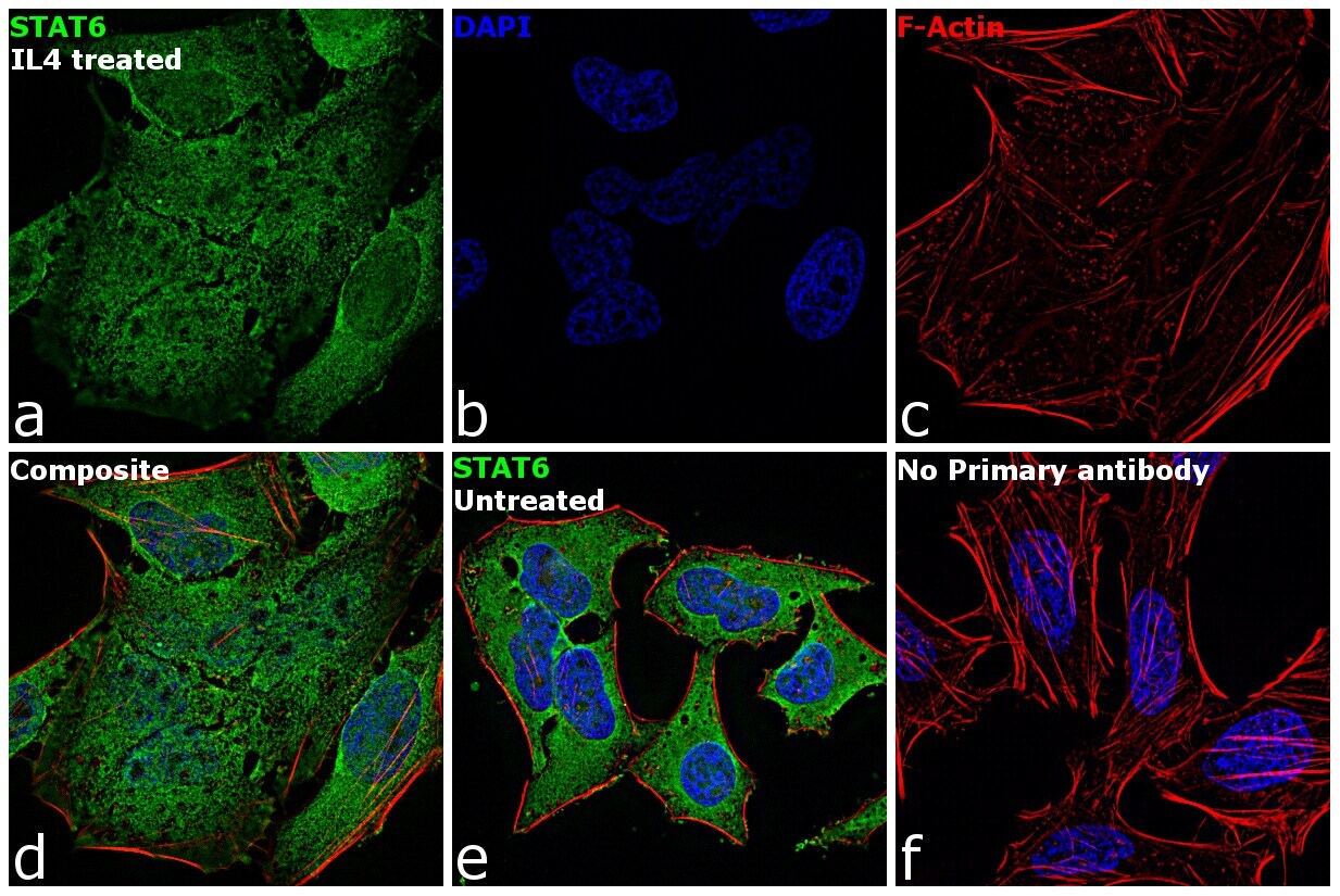
- Immunofluorescence analysis of STAT6 was performed using 70% confluent log phase HeLa cells treated with 10 ng of IL4 for 30 minutes. The cells were fixed with 4% paraformaldehyde for 10 minutes, permeabilized with 0.1% Triton™ X-100 for 15 minutes, and blocked with 1% BSA for 1 hour at room temperature. The cells were labeled with STAT6 Mouse Monoclonal Antibody (Product # MA5-15659) at 1:100 dilution in 0.1% BSA, incubated at 4 degree Celsius overnight and then labeled with Goat anti-Mouse IgG (H+L) Superclonal™ Secondary Antibody, Alexa Fluor® 488 conjugate (Product # A28175) at a dilution of 1:2000 for 45 minutes at room temperature (Panel a: green). Nuclei (Panel b: blue) were stained with SlowFade® Gold Antifade Mountant with DAPI (Product # S36938). F-actin (Panel c: red) was stained with Rhodamine Phalloidin (Product # R415, 1:300). Panel d represents the merged image showing cytoplasmic and nucleus localization. Panel e shows untreated cells with cytoplasmic signal. Panel f represents control cells with no primary antibody to assess background. The images were captured at 60X magnification.

- Submitted by
- Invitrogen Antibodies (provider)
- Main image

- Experimental details
- Immunofluorescence analysis of HeLa cells using STAT6 monoclonal antibody (Product # MA5-15659) (Green). Blue: DRAQ5 fluorescent DNA dye. Red: actin filaments have been labeled with phalloidin.

- Submitted by
- Invitrogen Antibodies (provider)
- Main image
- Experimental details
- Immunofluorescence analysis of HeLa cells using STAT6 monoclonal antibody (Product # MA5-15659) (Green). Blue: DRAQ5 fluorescent DNA dye. Red: actin filaments have been labeled with phalloidin.

- Submitted by
- Invitrogen Antibodies (provider)
- Main image

- Experimental details
- Immunofluorescence analysis of STAT6 was performed using 70% confluent log phase HeLa cells treated with 10 ng of IL4 for 30 minutes. The cells were fixed with 4% paraformaldehyde for 10 minutes, permeabilized with 0.1% Triton™ X-100 for 15 minutes, and blocked with 1% BSA for 1 hour at room temperature. The cells were labeled with STAT6 Mouse Monoclonal Antibody (Product # MA5-15659) at 1:100 dilution in 0.1% BSA, incubated at 4 degree Celsius overnight and then labeled with Goat anti-Mouse IgG (H+L) Superclonal™ Secondary Antibody, Alexa Fluor® 488 conjugate (Product # A28175) at a dilution of 1:2000 for 45 minutes at room temperature (Panel a: green). Nuclei (Panel b: blue) were stained with SlowFade® Gold Antifade Mountant with DAPI (Product # S36938). F-actin (Panel c: red) was stained with Rhodamine Phalloidin (Product # R415, 1:300). Panel d represents the merged image showing cytoplasmic and nucleus localization. Panel e shows untreated cells with cytoplasmic signal. Panel f represents control cells with no primary antibody to assess background. The images were captured at 60X magnification.
Supportive validation
- Submitted by
- Invitrogen Antibodies (provider)
- Main image
- Experimental details
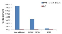
- Enrichment of endogenous STAT6 protein at specific gene loci using Anti-STAT6 Antibody: Chromatin Immunoprecipitation (ChIP) was performed using Anti-STAT6 Mouse Monoclonal Antibody (Product # MA5-15659, 8 µL) on sheared chromatin from 2 million HeLa cells starved overnight then treated with IL4 (100 ng/mL for 30 min) using the MAGnify ChIP system kit (Product # 49-2024). Normal Rabbit IgG was used as a negative IP control. The purified DNA was analyzed by qPCR with PCR primer pairs over DMD promoter, MS4A1 promoter (active) and SAT2 satellite repeats (inactive). Data is presented as fold enrichment of the antibody signal versus the negative control IgG using the comparative CT method.

- Submitted by
- Invitrogen Antibodies (provider)
- Main image

- Experimental details
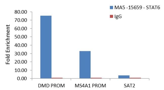
- Enrichment of endogenous STAT6 protein at specific gene loci using Anti-STAT6 Antibody: Chromatin Immunoprecipitation (ChIP) was performed using Anti-STAT6 Mouse Monoclonal Antibody (Product # MA5-15659, 8 µL) on sheared chromatin from 2 million HeLa cells starved overnight then treated with IL4 (100 ng/mL for 30 min) using the MAGnify ChIP system kit (Product # 49-2024). Normal Rabbit IgG was used as a negative IP control. The purified DNA was analyzed by qPCR with PCR primer pairs over DMD promoter, MS4A1 promoter (active) and SAT2 satellite repeats (inactive). Data is presented as fold enrichment of the antibody signal versus the negative control IgG using the comparative CT method.
Supportive validation
- Submitted by
- Invitrogen Antibodies (provider)
- Main image

- Experimental details
- NULL