Explore
 Validate
 Learn
Learn Western blot
Antibody data
- Antibody Data
- Antigen structure
- References [1]
- Comments [0]
- Validations
- Western blot [1]
- Flow cytometry [1]
Submit
Validation data
Reference
Comment
Report error
- Product number
- AF2167 - Provider product page

- Provider
- R&D Systems
- Product name
- Human/Mouse/Rat STAT6 aa 627-838 Antibody
- Antibody type
- Polyclonal
- Description
- Immunogen affinity purified. Detects human, mouse, and rat STAT6 in Western blots.
- Reactivity
- Human, Mouse, Rat
- Host
- Goat
- Conjugate
- Unconjugated
- Isotype
- IgG
- Vial size
- 100 ug
- Concentration
- LYOPH
- Storage
- Use a manual defrost freezer and avoid repeated freeze-thaw cycles. 12 months from date of receipt, -20 to -70 °C as supplied. 1 month, 2 to 8 °C under sterile conditions after reconstitution. 6 months, -20 to -70 °C under sterile conditions after reconstitution.
Submitted references Absence of Tumor Necrosis Factor Supports Alternative Activation of Macrophages in the Liver after Infection with Leishmania major.
Hu S, Marshall C, Darby J, Wei W, Lyons AB, Körner H
Frontiers in immunology 2018;9:1
Frontiers in immunology 2018;9:1
No comments: Submit comment
Supportive validation
- Submitted by
- R&D Systems (provider)
- Main image
- Experimental details
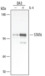
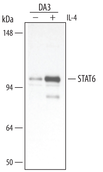
- Detection of Mouse STAT6 aa 627-838 by Western Blot. Western blot shows lysates of DA3 mouse myeloma cell line untreated (-) or treated (+) with 25 μg Recombinant Mouse IL-4 (Catalog # 404-ML) for 30 minutes. PVDF membrane was probed with 0.5 µg/mL of Goat Anti-Human/Mouse/Rat STAT6 Polyclonal Antibody (Catalog # AF2167, followed by HRP-conjugated Anti-Goat IgG Secondary Antibody (Catalog # HAF109). A specific band was detected for STAT6 aa 627-838 at approximately 100 - 110 kDa (as indicated). This experiment was conducted under reducing conditions and using Immunoblot Buffer Group 5.
Supportive validation
- Submitted by
- R&D Systems (provider)
- Main image

- Experimental details
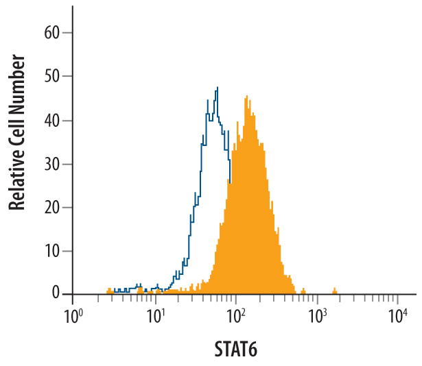
- Detection of STAT6 in HeLa Human Cell Line by Flow Cytometry. HeLa human cervical epithelial carcinoma cell line was stained with Goat Anti-Human/Mouse/Rat STAT6 aa 627-838 Antigen Affinity-purified Polyclonal Antibody (Catalog # AF2167, filled histogram) or control antibody (Catalog # AB-108-C, open histogram), followed by Allophycocyanin-conjugated Anti-Goat IgG Secondary Antibody (Catalog # F0108). To facilitate intracellular staining, cells were fixed with paraformaldehyde and permeabilized with methanol.