Explore
 Validate
 Learn
Learn Western blot
Antibody data
- Antibody Data
- Antigen structure
- References [1]
- Comments [0]
- Validations
- Western blot [2]
- Immunocytochemistry [2]
- Immunohistochemistry [2]
- Other assay [1]
Submit
Validation data
Reference
Comment
Report error
- Product number
- MA5-31488 - Provider product page

- Provider
- Invitrogen Antibodies
- Product name
- VAV1 Monoclonal Antibody (GT557)
- Antibody type
- Monoclonal
- Antigen
- Recombinant full-length protein
- Description
- Keep as concentrated solution. Predicted reactivity: Mouse (92%), Rat (92%), Pig (96%), Bovine (96%). Positive Control: Jurkat, NCI-H929. Store product as a concentrated solution. Centrifuge briefly prior to opening the vial.
- Reactivity
- Human, Mouse
- Host
- Mouse
- Isotype
- IgG
- Antibody clone number
- GT557
- Vial size
- 100 μL
- Concentration
- 1 mg/mL
- Storage
- Store at 4°C short term. For long term storage, store at -20°C, avoiding freeze/thaw cycles.
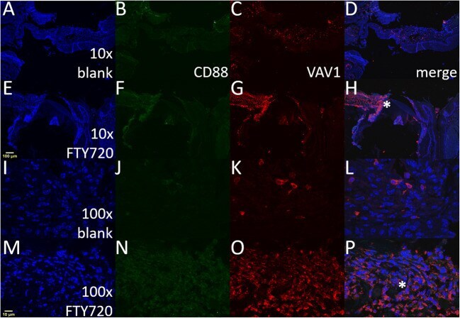
Submitted references Local delivery of FTY720 induces neutrophil activation through chemokine signaling in an oronasal fistula model.
Amanso AM, Turner TC, Kamalakar A, Ballestas SA, Hymel LA, Randall J, Johnston R, Arthur RA, Willett NJ, Botchwey EA, Goudy SL
Regenerative engineering and translational medicine 2021;7(2):160-174
Regenerative engineering and translational medicine 2021;7(2):160-174
No comments: Submit comment
Supportive validation
- Submitted by
- Invitrogen Antibodies (provider)
- Main image

- Experimental details
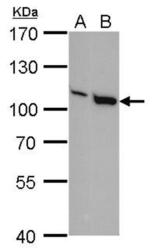
- VAV1 Monoclonal Antibody (GT557) detects VAV1 protein by Western blot analysis. A. 30 µg Jurkat whole cell lysate/extract. B. 30 µg NCI-H929 whole cell lysate/extract.7.5 % SDS-PAGE. VAV1 Monoclonal Antibody (GT557) (Product # MA5-31488) dilution: 1:1,000.

- Submitted by
- Invitrogen Antibodies (provider)
- Main image
- Experimental details
- Western blot analysis of VAV1 in A) Jurkat whole cell lysate, B) NCI-H929 whole cell lysate using VAV1 monoclonal antibody (Product # MA5-31488) using 30 µg of sample at a dilution of 1:1000. Prior to incubation with primary antibody, the sample was separated on 7.5% SDS-PAGE.

Supportive validation
- Submitted by
- Invitrogen Antibodies (provider)
- Main image
- Experimental details
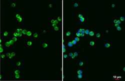
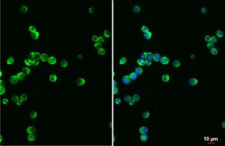
- Immunocytochemistry-Immunofluorescence analysis of VAV1 was performed in Jurkat cells fixed in 4% paraformaldehyde at RT for 15 min. Green: VAV1 Monoclonal Antibody (GT557) (Product # MA5-31488) diluted at 1:500. Blue: Fluoroshield with DAPI. Scale bar = 10 µm.
- Submitted by
- Invitrogen Antibodies (provider)
- Main image
- Experimental details
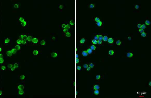
- Immunocytochemistry-Immunofluorescence analysis of VAV1 was performed in Jurkat cells fixed in 4% paraformaldehyde at RT for 15 min. Green: VAV1 Monoclonal Antibody (GT557) (Product # MA5-31488) diluted at 1:500. Blue: Fluoroshield with DAPI. Scale bar = 10 µm.

Supportive validation
- Submitted by
- Invitrogen Antibodies (provider)
- Main image

- Experimental details
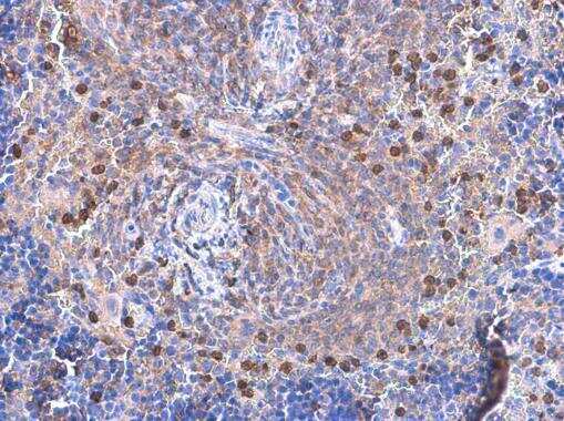
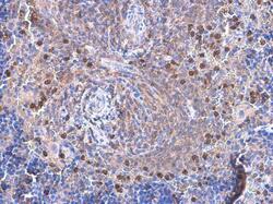
- Immunohistochemistry (Paraffin) analysis of VAV1 was performed in paraffin-embedded mouse spleen tissue using VAV1 Monoclonal Antibody (GT557) (Product # MA5-31488) at a dilution of 1:200.
- Submitted by
- Invitrogen Antibodies (provider)
- Main image
- Experimental details
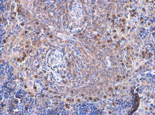
- Immunohistochemistry (Paraffin) analysis of VAV1 was performed in paraffin-embedded mouse spleen tissue using VAV1 Monoclonal Antibody (GT557) (Product # MA5-31488) at a dilution of 1:200.
Supportive validation
- Submitted by
- Invitrogen Antibodies (provider)
- Main image
- Experimental details
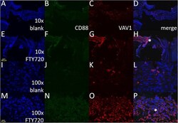
- Fig. 5 FTY720 induces protein expression of Vav1 at the site of the wound healing after palatal injury. Representative immunofluorescence images of mice palates 3 days post-injury stained against two important immunomodulators, Vav1 and CD88 proteins. (a-d) and (i-l) panels are from injured palate treated with blank nanofiber scaffold (vehicle). (e-h) and (m-p) panels are from injured palate treated with FTY720 nanofiber scaffold. The panels (a, e, i, and m) are nuclear staining using DAPI (blue). Panels (b, f, g, and n) are staining against CD88-Alexa488 (green). Panels (c, g, k, and o) are staining against Vav1-Alexa568 (red). Panels (d, h, l, and p) are the merged images. The asterisk symbol signalizes the same region in the injured palate treated with FTY720 between x10 (H) and x100 (P) panels. The images were acquired using the x10 and x100 objective on an Olympus F1000 microscope. The scale bars are noted in yellow, 10 mum for x10 (A) and 100 mum for x100 (M) images