Explore
 Validate
 Learn
Learn Western blot
Antibody data
- Antibody Data
- Antigen structure
- References [1]
- Comments [0]
- Validations
- Western blot [1]
- Immunocytochemistry [1]
- Other assay [1]
Submit
Validation data
Reference
Comment
Report error
- Product number
- MA1-24966 - Provider product page

- Provider
- Invitrogen Antibodies
- Product name
- beta Amyloid Monoclonal Antibody (DE2B4)
- Antibody type
- Monoclonal
- Antigen
- Synthetic peptide
- Description
- Protein digestion pretreatment of paraffin sections using formic acid for 2 - 3 minutes is required prior to staining with MA1-24966.
- Antibody clone number
- DE2B4
- Concentration
- 1 mg/mL
Submitted references Beta-amyloid induces apoptosis of neuronal cells by inhibition of the Arg/N-end rule pathway proteolytic activity.
Kechko OI, Petrushanko IY, Brower CS, Adzhubei AA, Moskalev AA, Piatkov KI, Mitkevich VA, Makarov AA
Aging 2019 Aug 24;11(16):6134-6152
Aging 2019 Aug 24;11(16):6134-6152
No comments: Submit comment
Supportive validation
- Submitted by
- Invitrogen Antibodies (provider)
- Main image
- Experimental details
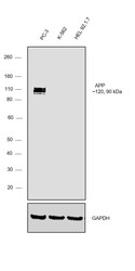
- Western blot was performed using Anti-beta Amyloid Monoclonal Antibody (DE2B4)(Product # MA1-24966) and a 120, 90kDa band corresponding to beta Amyloid was observed in PC-3 and not in K-562 and HEL 92.1.7. Whole cell extracts (30 µg lysate) of PC-3 (Lane 1), K-562 (Lane 2) and HEL 92.1.7 (Lane 3), were electrophoresed using NuPAGE™ 4-12% Bis-Tris Protein Gel (Product # NP0321BOX). Resolved proteins were then transferred onto a Nitrocellulose membrane (Product # IB23001) by iBlot® 2 Dry Blotting System (Product # IB21001). The blot was probed with the primary antibody (2ug/ml) and detected by chemiluminescence with Goat anti-Mouse IgG (H+L) Superclonal™ Recombinant Secondary Antibody, HRP (Product # A28177,1:4000 dilution) using the iBright FL 1000 (Product # A32752). Chemiluminescent detection was performed using Novex® ECL Chemiluminescent Substrate Reagent Kit (Product # WP20005).
Supportive validation
- Submitted by
- Invitrogen Antibodies (provider)
- Main image
- Experimental details
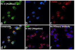
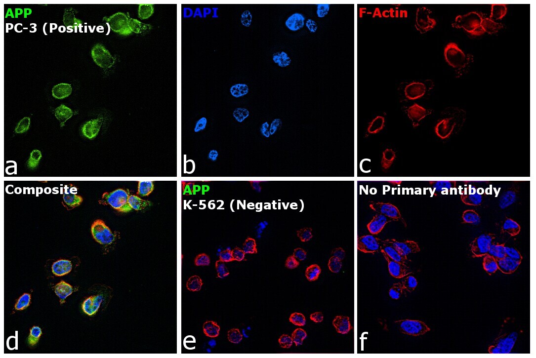
- Immunofluorescence analysis of beta Amyloid was performed using 70% confluent log phase PC-3 cells. The cells were fixed with 4% paraformaldehyde for 10 minutes, permeabilized with 0.1% Triton™ X-100 for 15 minutes, and blocked with 2% BSA for 45 minutes at room temperature. The cells were labeled with beta Amyloid Monoclonal Antibody (DE2B4) (Product # MA1-24966) at 1:200 dilution in 0.1% BSA, incubated at 4 degree celsius overnight and then labeled with Goat anti-Mouse IgG (H+L) Highly Cross-Adsorbed Secondary Antibody, Alexa Fluor Plus 488 (Product # A32723), (1:3000 dilution), for 45 minutes at room temperature (Panel a: Green). Nuclei (Panel b: Blue) were stained with ProLong™ Diamond Antifade Mountant with DAPI (Product # P36962). F-actin (Panel c: Red) was stained with Rhodamine Phalloidin (Product # R415, 1:300). Panel d represents the merged image showing Cytoplasm, Plasma membrane and Nuclear localization. Panel e represents merged image for K-562 cells showing no staining for APP. Panel f represents control cells with no primary antibody to assess background. The images were captured at 60X magnification.
Supportive validation
- Submitted by
- Invitrogen Antibodies (provider)
- Main image
- Experimental details
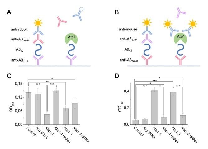
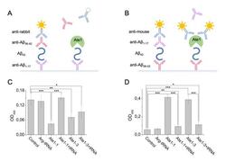
- Figure 3 Interaction of Abeta 42 , tRNA, and Ate1. Schematic representation of ELISA assay with immobilized anti-Abeta1-17 antibodies ( A ) or anti-Abeta36-42 antibodies ( B ). ( C ) Detection of tRNA and Ate1 ability to interact with C-terminus of Abeta. ( D ) Same as (C) but with N-terminal region of Abeta. OD 450 - optical density measured at 450 nm. Each value is the mean +- SD of at least four independent experiments; *p < 0.04, **p < 0.01, ***p < 0.001.