Explore
 Validate
 Learn
Learn Western blot
 Immunohistochemistry
Antibody data
- Antibody Data
- Antigen structure
- References [5]
- Comments [0]
- Validations
- Western blot [2]
- Immunocytochemistry [1]
Submit
Validation data
Reference
Comment
Report error
- Product number
- MA1-25489 - Provider product page

- Provider
- Invitrogen Antibodies
- Product name
- Amyloid Precursor Protein Monoclonal Antibody (3E9)
- Antibody type
- Monoclonal
- Antigen
- Synthetic peptide
- Description
- Store product as a concentrated solution. Centrifuge briefly prior to opening the vial.
- Reactivity
- Human, Mouse
- Host
- Mouse
- Isotype
- IgG
- Antibody clone number
- 3E9
- Vial size
- 50 µg
- Concentration
- 1 mg/mL
- Storage
- Store at 4°C short term. For long term storage, store at -20°C, avoiding freeze/thaw cycles.
Submitted references Limiting Neuronal Nogo Receptor 1 Signaling during Experimental Autoimmune Encephalomyelitis Preserves Axonal Transport and Abrogates Inflammatory Demyelination.
Nogo-receptor 1 deficiency has no influence on immune cell repertoire or function during experimental autoimmune encephalomyelitis.
Ran-independent nuclear import of cyclin B1-Cdc2 by importin beta.
Ran-independent nuclear import of cyclin B1-Cdc2 by importin beta.
Sequence and characterization of cytoplasmic nuclear protein import factor p97.
Lee JY, Kim MJ, Thomas S, Oorschot V, Ramm G, Aui PM, Sekine Y, Deliyanti D, Wilkinson-Berka J, Niego B, Harvey AR, Theotokis P, McLean C, Strittmatter SM, Petratos S
The Journal of neuroscience : the official journal of the Society for Neuroscience 2019 Jul 10;39(28):5562-5580
The Journal of neuroscience : the official journal of the Society for Neuroscience 2019 Jul 10;39(28):5562-5580
Nogo-receptor 1 deficiency has no influence on immune cell repertoire or function during experimental autoimmune encephalomyelitis.
Litwak SA, Payne NL, Campanale N, Ozturk E, Lee JY, Petratos S, Siatskas C, Bakhuraysah M, Bernard CC
PloS one 2013;8(12):e82101
PloS one 2013;8(12):e82101
Ran-independent nuclear import of cyclin B1-Cdc2 by importin beta.
Takizawa CG, Weis K, Morgan DO
Proceedings of the National Academy of Sciences of the United States of America 1999 Jul 6;96(14):7938-43
Proceedings of the National Academy of Sciences of the United States of America 1999 Jul 6;96(14):7938-43
Sequence and characterization of cytoplasmic nuclear protein import factor p97.
Chi NC, Adam EJ, Adam SA
The Journal of cell biology 1995 Jul;130(2):265-74
The Journal of cell biology 1995 Jul;130(2):265-74
No comments: Submit comment
Supportive validation
- Submitted by
- Invitrogen Antibodies (provider)
- Main image

- Experimental details
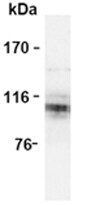
- Western blot analysis of Amyloid Precursor Protein expression in mouse brain using Amyloid Precursor Protein Monoclonal Antibody (3E9) (Product # MA1-25489).
- Submitted by
- Invitrogen Antibodies (provider)
- Main image
- Experimental details
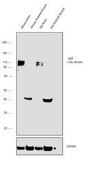
- Western blot was performed using Anti-Amyloid Precursor Protein Monoclonal Antibody (3E9)(Product # MA1-25489) and a 120, 90kDa band corresponding to Amyloid Precursor Protein was observed across the panel tested except for Mouse Skeletal Muscle and Rat Skeletal Muscle which are reported to be negative. Whole cell extracts (30 µg lysate) of Mouse Brain (Lane 1), Mouse Skeletal Muscle (Lane 2), Rat Brain (Lane 3) and Rat Skeletal Muscle (Lane 4), were electrophoresed using NuPAGE™ 4-12% Bis-Tris Protein Gel (Product # NP0321BOX). Resolved proteins were then transferred onto a Nitrocellulose membrane (Product # IB23001) by iBlot® 2 Dry Blotting System (Product # IB21001). The blot was probed with the primary antibody (5ug/ml) and detected by chemiluminescence with Goat anti-Mouse IgG (H+L) Superclonal™ Recombinant Secondary Antibody, HRP (Product # A28177,1:4000 dilution) using the iBright FL 1000 (Product # A32752). Chemiluminescent detection was performed using Novex® ECL Chemiluminescent Substrate Reagent Kit (Product # WP20005).

Supportive validation
- Submitted by
- Invitrogen Antibodies (provider)
- Main image
- Experimental details
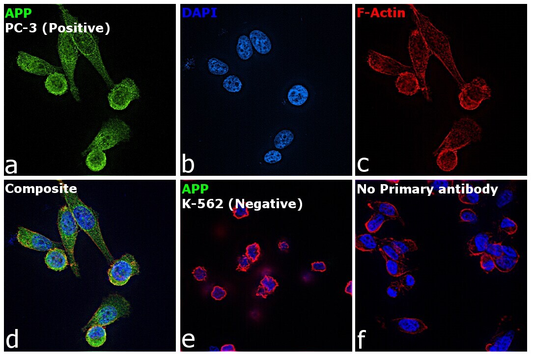
- Immunofluorescence analysis of Amyloid Precursor Protein was performed using 70% confluent log phase PC-3 cells. The cells were fixed with 4% paraformaldehyde for 10 minutes, permeabilized with 0.1% Triton™ X-100 for 15 minutes, and blocked with 2% BSA for 45 minutes at room temperature. The cells were labeled with Amyloid Precursor Protein Monoclonal Antibody (3E9) (Product # MA1-25489) at 1:200 in 0.1% BSA, incubated at 4 degree celsius overnight and then labeled with Goat anti-Mouse IgG (H+L) Highly Cross-Adsorbed Secondary Antibody, Alexa Fluor Plus 488 (Product # A32723), (1:3000 dilution), for 45 minutes at room temperature (Panel a: Green). Nuclei (Panel b: Blue) were stained with ProLong™ Diamond Antifade Mountant with DAPI (Product # P36962). F-actin (Panel c: Red) was stained with Rhodamine Phalloidin (Product # R415, 1:300 dilution). Panel d represents the merged image showing Cytoplasm, Plasma membrane and Nuclear localization. Panel e represents merged image for K-562 cells showing no staining for APP. Panel f represents control cells with no primary antibody to assess background. The images were captured at 60X magnification.