Explore
 Validate
 Learn
Learn Western blot
 Immunohistochemistry
Antibody data
- Antibody Data
- Antigen structure
- References [2]
- Comments [0]
- Validations
- Immunohistochemistry [1]
- Other assay [1]
Submit
Validation data
Reference
Comment
Report error
- Product number
- PA1-84165 - Provider product page

- Provider
- Invitrogen Antibodies
- Product name
- Amyloid Precursor Protein Polyclonal Antibody
- Antibody type
- Polyclonal
- Antigen
- Synthetic peptide
- Description
- PA1-84165 recognizes both intact APP and also the C99 fragment generated by Beta-secretase. The sequence recognized by this antibody corresponds to amino acids 85-99 of the C99 fragment. Heat-mediated antigen retrieval is recommended for the staining of paraffin sections.
- Reactivity
- Human, Mouse, Rat
- Host
- Rabbit
- Isotype
- IgG
- Vial size
- 100 µg
- Concentration
- 1.0 mg/mL
- Storage
- Store at 4°C short term. For long term storage, store at -20°C, avoiding freeze/thaw cycles.
Submitted references Depletion of microglia immediately following traumatic brain injury in the pediatric rat: Implications for cellular and behavioral pathology.
Alzheimer's disease related markers, cellular toxicity and behavioral deficits induced six weeks after oligomeric amyloid-β peptide injection in rats.
Hanlon LA, Raghupathi R, Huh JW
Experimental neurology 2019 Jun;316:39-51
Experimental neurology 2019 Jun;316:39-51
Alzheimer's disease related markers, cellular toxicity and behavioral deficits induced six weeks after oligomeric amyloid-β peptide injection in rats.
Zussy C, Brureau A, Keller E, Marchal S, Blayo C, Delair B, Ixart G, Maurice T, Givalois L
PloS one 2013;8(1):e53117
PloS one 2013;8(1):e53117
No comments: Submit comment
Supportive validation
- Submitted by
- Invitrogen Antibodies (provider)
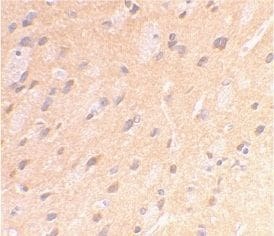
- Main image
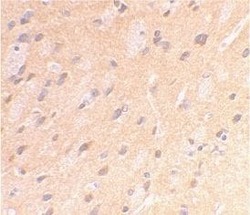
- Experimental details
- Immunohistochemical staining of rat brain using a Amyloid Precursor Protein polyclonal antibody (Product # PA1-84165)
Supportive validation
- Submitted by
- Invitrogen Antibodies (provider)
- Main image

- Experimental details
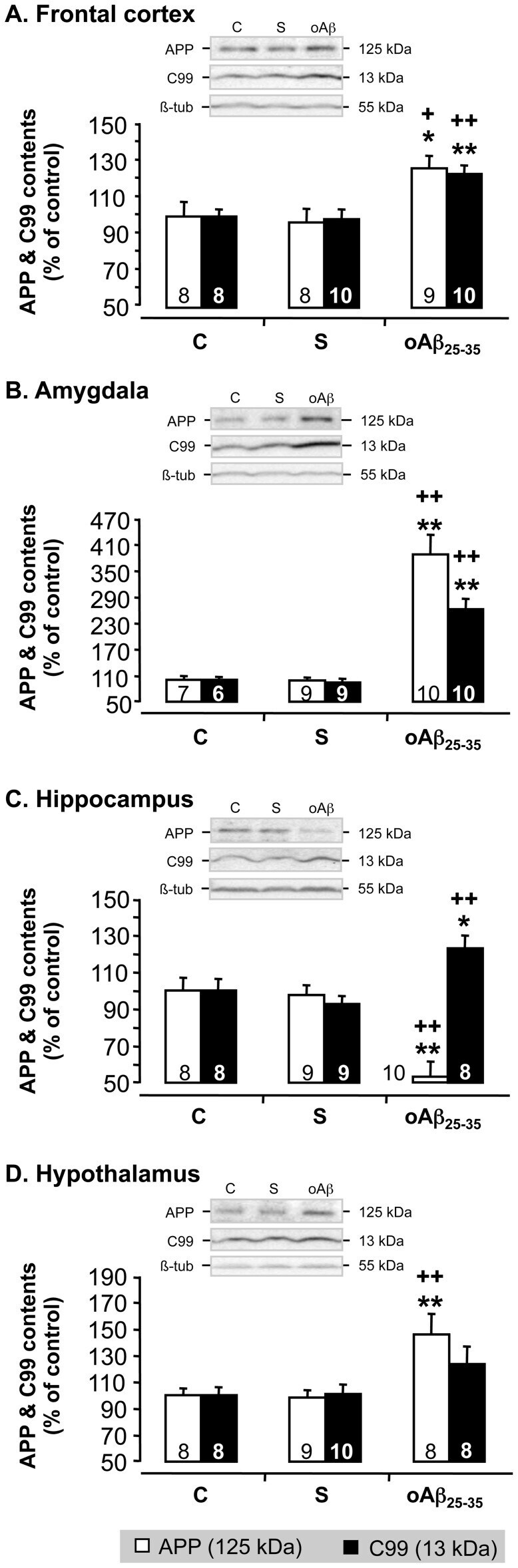
- Figure 12 APP processing. Effects of oAbeta 25-35 (10 ug/rat) icv injection on APP processing in the frontal cortex, amygdala, hippocampus and hypothalamus, determined by western blot in untreated control rats and 6 weeks after Abeta 25-35 scrambled amyloid peptide (10 ug/rat; negative control) or oAbeta 25-35 injection. APP (125 KDa) and C99 (13 KDa) variations were normalized with beta-tubulin (beta-tub, 55 KDa) variations and compared with non-injected rats (control group: C). The results are expressed as means +- SEM. *p